38 Approach to Pediatric Electromyography
Neuromuscular Diagnoses Are Different in Children Than in Adults
The most common referral diagnoses to the typical electromyography (EMG) laboratory include radiculopathy, polyneuropathy, and carpal tunnel syndrome. However, adults are more commonly studied in the EMG laboratory, so this group of diagnoses reflects neuromuscular conditions seen in the adult age group. In contrast, the neuromuscular disorders seen in children often are different. For example, entrapment neuropathies are very common in adults but are extremely rare in children. Likewise, radiculopathy, probably the most common of all EMG referral diagnoses, is virtually unheard of in children, except in cases of trauma. Although peripheral neuropathies occur in children, they are most often genetic, whereas most peripheral neuropathies in adults referred to the EMG laboratory are acquired disorders, usually toxic, metabolic, inflammatory, or associated with other coexistent medical illnesses. Unlike adults, the more common diagnoses in children referred to the EMG laboratory are inherited disorders of the motor unit, including the anterior horn cell (e.g., spinal muscular atrophy), peripheral nerve (e.g., Charcot–Marie–Tooth), or muscle (e.g., muscular dystrophy).
Children with neuromuscular disorders often present clinically as a delay in motor milestones. In many cases, it may not be clear from the symptoms and signs whether the etiology is central or peripheral. One of the best examples of this predicament is that of the floppy infant, in whom the differential diagnosis includes the entire length of the neuraxis, from brain to muscle. In this regard, EDX studies often are helpful in differentiating peripheral from central etiologies and, accordingly, guiding the subsequent evaluation in a useful and logical direction.
Maturation Issues
When studying children, it is essential to appreciate what is normal for what age. This is especially important when interpreting conduction velocities and differentiating a normal conduction velocity from axonal loss or demyelination. Most adult electromyographers who study adults are well versed in the EDX criteria for demyelination:
• Conduction velocities less than 75% the lower limit of normal
• Distal latencies and late responses greater than 130% the upper limit of normal
• Conduction block, which signifies not only demyelination but acquired demyelination
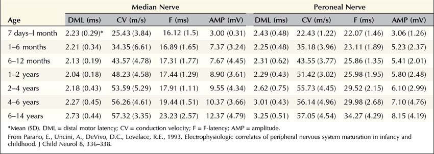
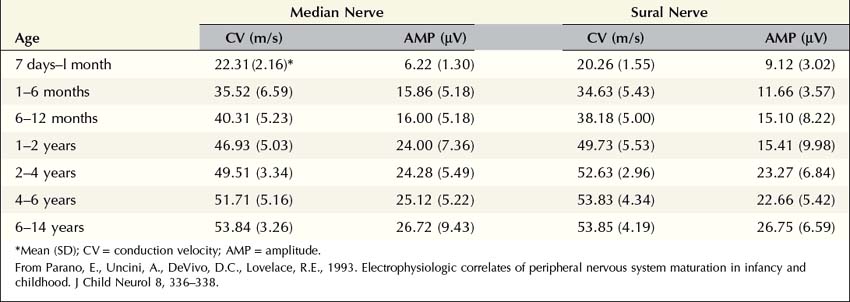
However, infants and young children often have slowed conduction velocities that would be considered in the “demyelinating range” for adults. In most cases, this is not because infants and young children have demyelinated nerves; rather, they have nerves that have yet to be myelinated in the first place. The process of myelination is age dependent, beginning in utero, with nerve conduction velocities in full-term infants approximately half that of adult normal values. Accordingly, nerve conduction velocities of 25 to 30 m/s are normal at birth. Conduction velocity rapidly increases after birth, reaching approximately 75% of adult normal values by age 1 year, and the adult range by age 3 to 5 years, when myelination is complete. Accordingly, when a child is studied in the EMG laboratory, it is essential that age-based normal control values are used (Tables 38–1 and 38–2).
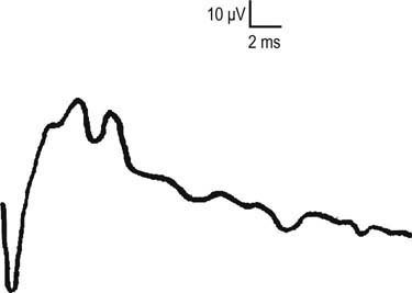
One interesting aspect of myelin maturation is often observed during the nerve conduction studies. Many are familiar with the fact that different white matter tracts in the central nervous system myelinate at different times. Indeed, one can often use the pattern of myelination on a brain magnetic resonance imaging (MRI) scan to correctly predict the age of a young child. Similarly, different fibers in the peripheral nervous system myelinate at different times as well. In the EMG laboratory, this often manifests as a bifid morphology (i.e., two separate peaks) on sensory nerve action potentials (SNAPs) in infants and children (Figure 38–1). This bifid morphology is due to some fibers having already been fully myelinated (the first peak), whereas others have not and trail behind (i.e., the second peak). It is not unusual to see bifid SNAPs between the ages of 3 months and 4 to 6 years. These bifid SNAPs are a completely normal finding. Eventually, as the fibers in the second peak fully myelinate, the second peak moves to the left and merges with the first peak. This forms a larger sensory response, as is typically seen in adults.

FIGURE 38–1 Sural sensory nerve action potential in a young child.
Note the bifid morphology. These bifid sensory responses are a completely normal finding between the ages of 3 months through 4 to 6 years. They occur as different populations of fibers myelinate at different times. Eventually, the group of fibers in the second peak will fully myelinate. The second peak will move to the left and merge with the first peak to form a larger sensory response.
As with adults, the F response can be easily studied in children. Although the F response often is thought of as evaluating the proximal nerve segments, it assesses the entire length of the nerve, from the stimulation point to the spinal cord and back, and then past the stimulation point to the muscle. Thus, the F response latency depends not only on the conduction velocity and distal latency but also on the length of the limb. Because infants and children have slower conduction velocities than adults, one would expect the F responses to be very long. However, counterbalancing this is the very short limb length of a child compared to an adult. Thus, there are two opposing influences on the F response in children: limb length and conduction velocity. In infants and young children, the influence of the limb length is more overriding, resulting in F-wave latencies that are much shorter in children than adults (typically in the range of 16–19 ms in the upper extremities). Thus, whenever an F response is performed on a child, it is essential to compare it to normal control values for the child’s age or height.
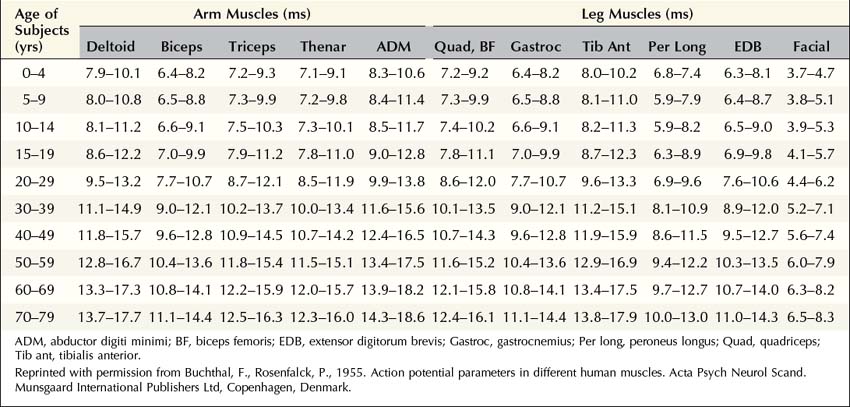
The most important maturation issue for the needle EMG portion of the examination is the size of the motor unit. It is no surprise that the physical size of a motor unit of a newborn is much smaller than that of an adult. Transverse motor unit territory increases greatly with age, doubling from birth to adulthood, mostly because of the increase in individual muscle fiber size. Thus, normal motor unit action potentials (MUAPs) in infants typically are very small, representing the physical size of the motor unit. Indeed, in infants, it often is difficult to differentiate normal MUAPs from myopathic ones. This once again underscores that when one interprets EDX findings in children, including MUAPs, it is essential to use age-based normal control values (Table 38–3).
Technical Issues
A large number of unique technical issues must be kept in mind when studying infants and children so that reliable and accurate data can be obtained. The first important issue is measurement of distances and its relationship to technical errors. Because a child’s limb is much smaller than an adult’s, much shorter distances are used. When short distances are used, a small error in measurement creates a much larger error in computed conduction velocities than when longer distances are used. For instance, in an adult, if the distance between the wrist and elbow is measured at 20 cm but is off by 1 cm (i.e., the true measurement is 21 cm), this results in an error of 5% when calculating a conduction velocity. However, in a newborn baby, if the measured distance is 7 cm but is off by 1 cm (i.e., the true measurement is 8 cm), the error in conduction velocity increases to 14%. Thus, one needs to be especially careful when measuring distances in children.
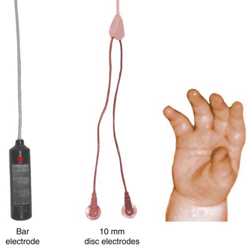
Second, smaller electrodes often are needed in infants and young children because their limbs and muscles are so small. The typical bar electrode that has the active and reference contacts separated by 2.5 cm often is too large for most infants and small children (Figure 38–2). Standard 10 mm disc electrodes often will suffice for most ages, except for newborns in which smaller electrodes generally are needed. Likewise, the standard adult stimulator often is too large for infants and young children because of the size of the prongs and the distance between the cathode and anode. Often it is preferable to use a pediatric-sized stimulator so that the nerve of interest is more accurately stimulated (Figure 38–3).

FIGURE 38–2 Pediatric electrodiagnostic studies and recording electrode size.
The standard bar electrode (left) and 10 mm disc electrodes (middle) are compared to the size of an infant’s hand (right). Smaller electrodes may be needed in infants and young children because their limbs and muscles are so small. Standard 10 mm disc electrodes often will suffice for most age groups, including infants. In newborns, however, smaller electrodes are needed. Other standard electrodes, like the bar electrode, are too large for a newborn or infant’s hand.

Stay updated, free articles. Join our Telegram channel

Full access? Get Clinical Tree

