Figure 60-1 A, Photomicrograph of sural nerve biopsy, paraffin-embedded, cross-section stained with hematoxylin and eosin. There is a prominent subperineurial, hypercellular focus within one of the nerve fascicles. B, Photomicrograph of sural nerve biopsy, paraffin-embedded, cross-section, adjacent to section presented at left, immunostained for epithelial membrane antigen. There is positive immunostaining of the subperineurial hypercellular focus, which is consistent with an intraneural perineurioma. The non-neoplastic perineurial cells of the perineurium also immunostain for epithelial membrane antigen.
(This photomicrograph is kindly provided by Dr. Peter Dyck and the Mayo Peripheral Nerve Center).
Biopsy of the right optic nerve reveals an optic nerve sheath meningioma that compresses the contiguous optic nerve (Fig. 60-2A). The meningioma has numerous psammoma bodies and cellular whorls (see Fig. 60-2B).
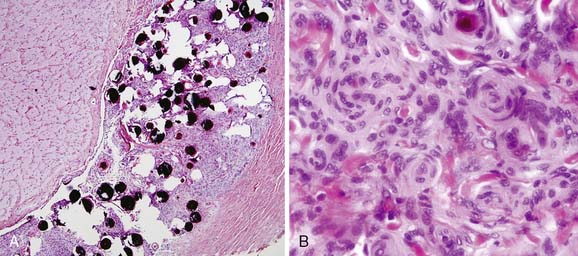
Figure 60-2 A, Photomicrograph of optic nerve biopsy, paraffin-embedded, cross-section, stained with hematoxylin and eosin. There is atrophic optic nerve in the left third of the image, meningioma with prominent psammoma bodies in the middle third of the image, and the optic nerve’s dural sheath in the lower right third of the image. B, Photomicrograph of optic nerve biopsy, paraffin-embedded, cross-section stained with hematoxylin and eosin. Higher magnification of the meningioma shows the characteristic cellular whorls within the neoplasm.
CONCLUSIONS
An association between peripheral neuropathy and multiple cranial neuropathies is unusual, although it can be seen in both inherited (hereditary motor sensory neuropathy types III, VI) and acquired (chronic inflammatory demyelinating polyneuropathy, sarcoidosis, diabetes) peripheral neuropathies. Intraneural perineurioma is a rare, benign, nerve sheath tumor that typically presents with isolated peripheral mononeuropathy. Only rarely does intraneural perineurioma present as a cranial neuropathy.1 Intracranial meningioma may rarely be associated with cranial mononeuropathies.2 To our knowledge, prior cases of multiple peripheral and cranial mononeuropathies, with pathologic evidence of perineurioma in peripheral nerve and meningioma in cranial nerve, have not been reported. The association of intraneural perineurioma and meningioma may be more than coincidental. Both neoplasms have been associated with chromosome 22 abnormalities.1,3,4 There has been no clear association in the literature between intraneural perineurioma and neurofibromatosis.1,5 Previously reported cases of perineurioma-associated polyneuropathy with multiple cranial mononeuropathies only documented the intraneural perineurioma of peripheral nerve; the cause of the cranial mononeuropathies was not established.5–7 Without additional cranial nerve biopsies, the cause of the remaining cranial mononeuropathies in our case remains to be determined. The relationship between tumors arising from different cell types in the central and peripheral nervous systems raises the intriguing question as to why in this particular patient this association has occurred and why it is not an association seen more often clinically.
Related posts:
 Malignant Peripheral Nerve Sheath Tumor
Disseminated Sporotrichosis with Multiple Granulomatous Mononeuropathies
Late Sporadic CMT4C—A New KIAA1985 Mutation
Late-Onset Transthyretin Val30Met Familial Amyloid Polyneuropathy Unrelated to Endemic Foci
A Weak, and Numb Patient with Tremor—Antimyelin-Associated Glycoprotein Polyneuropathy
Copper Deficiency Myeloneuropathy
Malignant Peripheral Nerve Sheath Tumor
Disseminated Sporotrichosis with Multiple Granulomatous Mononeuropathies
Late Sporadic CMT4C—A New KIAA1985 Mutation
Late-Onset Transthyretin Val30Met Familial Amyloid Polyneuropathy Unrelated to Endemic Foci
A Weak, and Numb Patient with Tremor—Antimyelin-Associated Glycoprotein Polyneuropathy
Copper Deficiency Myeloneuropathy
Stay updated, free articles. Join our Telegram channel

Full access? Get Clinical Tree


