NASCET degree of stenosis. The distal diameter “D” is the denominator:
Degree = (1 − R/D) × 100%.
It can be seen that this results in a lower degree of stenosis compared to the ECST degree of stenosis using the local diameter as denominator.
The local diameter reduction describes better the plaque burden. Any report of ultrasonic results describing degree of stenosis in percent has to make clear to what type of radiologic grading this is referring: NASCET or ECST. A concentric type of stenosis leads to more area reduction and higher velocities than an eccentric one [3]. This limits the estimation of the hemodynamic effect based on diameter reduction.
Hemodynamic effects
Any normal or abnormal vascular condition resulting in changes in caliber, bending, branching or circumscribed wall thickening induces changes of local flow characteristics. A severe stenosis or occlusion induces additional effects in the up- and downstream vascular territory due to a pressure drop, reduced flow volume and formation of collateral flow. In the context of grading a stenosis, the definition of the term “hemodynamic effect” should be limited to changes of the flow pattern distal to the stenosis. The hemodynamic effect characterizes a severe stenosis. We separate severe from moderate stenoses based on the existence of “hemodynamic effect.” But it is essential to take into consideration the whole cerebral circulation including the contralateral vessels, the anterior and posterior circulation and the existence of collateral pathways in order to make a correct interpretation of local and distal effects on flow velocities. The existence and capacity or absence of collateral flow (complete or incomplete circle of Willis) strongly influences the velocities in the stenosis [4]. This is an argument for the proposed multiparametric diagnostic approach (Table 5C.1) as explained in detail in the following chapters.
| Degree of stenosis as defined by NASCET (%) | Grading of internal carotid stenosis | ||||||
|---|---|---|---|---|---|---|---|
| 10–40 | 50 | 60 | 70 | 80 | 90 | Occlusion | |
| Main criteria | |||||||
| 1. B-mode image, diameter | Applicable | Possibly applicable | Imaging of occluded artery | ||||
| 2. Color Doppler image | Plaque delineation | Flow | Flow | Flow | Flow | Flow | Absence of flow |
| 3. PSV threshold (cm/s) | 125 | 230 | NA | NA | |||
| 4a. PSV average (cm/s) | ≤160 | 210 | 240 | 330 | 370 | Variable | NA |
| 4b. PSV poststenotic (cm/s) | ≥50 | <50 | <30 | NA | |||
| 5. Collateral flow (periorbital arteries or circle of Willis) | Possible | Present | Present | Present | |||
| Additional criteria | |||||||
| 6. Prestenotic flow (diastole) (CCA) | Possibly reduced | Reduced | Reduced | Reduced | |||
| 7. Poststenotic flow disturbances (severity and length) | Moderate | Pronounced | Pronounced | Pronounced | Variable | NA | |
| 8. End-diastolic flow velocity in the stenosis (cm/s) | <100 | >100 | Variable | NA | |||
| 9. Carotid radio ICA/CCA | <2 | ≥2 | ≥2 | >4 | >4 | Variable | NA |
CCA, common carotid artery; ICA, internal carotid artery; NA, not applicable; PSV, peak systolic velocity.
Velocities in a stenosis
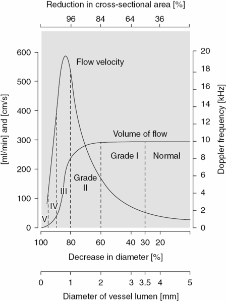
The relationship between the stenotic lumen and flow velocity in stenosis and flow volume has been described in the key paper by Spencer and Reid [3] (Figure 5C.2). Flow volume is stable over a broad range of narrowing while velocities in the stenosis are increasing. With a high degree of stenosis, flow volume starts to decrease while velocities still increase. Finally, approaching occlusion, both flow volume and flow velocities will decrease. Therefore, the peak systolic velocity (PSV, e.g., 200 cm/s) as a unique criterion is not enough to decide whether the stenosis is moderate, severe or near to occlusion. Other findings help to differentiate between these situations. Only with a nearly occluded artery, is poststenotic flow velocity (as volume flow) clearly diminished and signs of the hemodynamic effect are very clear: reduced velocity in the prestenotic segment and established collateral flow. With a moderate stenosis this is definitively not the case. Velocities in a stenosis are strongly influenced by collateral flow. Velocities may increase by contralateral ICA occlusion [5]. Less known is that velocities increase when there is a low collateral flow rate to the territory supplied by the stenosed artery due to an incomplete circle of Willis. This leads to a high poststenotic pressure drop, driving velocities up. The opposite is the case with good collateral flow through the circle of Willis: velocities are relatively lower in this case due to a higher poststenotic pressure and with lower pressure drop. Spencer demonstrated in a model that this effect may account for huge variations in velocities with the same morphologic degree of stenosis [4].

Theoretical relationship between flow velocity and volume of flow for different degrees of stenosis. Doppler frequencies based on continuous-wave Doppler, 4-MHz emission frequency.
Ultrasonic modalities
B-mode imaging of morphology
With modern techniques using high-frequency scan heads, the vessel wall can be shown with high resolution and characterized by its echogenicity. Vessels can be imaged with longitudinal and cross-sectional planes. Low-grade disease is well demonstrated by this ultrasonic modality. With increasing degree of stenosis and arteriosclerotic remodeling of the vessel wall, the B-mode image is more and more influenced by artifacts (e.g., reverberations or shadowing due to calcifications). This is why B-mode imaging is essential for localizing and characterizing moderate to severe disease but additional hemodynamic parameters become more important with severe stenosis.
Color imaging and flow field
The distribution of flow over the vessel diameter can be shown by color flow imaging. With adequate setting of the “color window” (pulse repetition frequency, PRF) it is possible to show the directional flow lines as well as a local increase of velocities due to the stenosis and disturbed flow. This method, however, should not be used to measure diameter or area reduction. Color imaging is highly dependent on gain and PRF. In addition, the frame rate of color images is slower than that of B-mode imaging leading to smearing of color over the vessel edges due to longitudinal and cross-sectional vessel pulsations. Color imaging is therefore predominately serving as a pathfinder, that is, where to best measure velocities by means of the pulsed Doppler rather than being used for grading a stenosis. On the other hand, color is used to delineate plaques of low echogenicity and is essential in differentiating occlusion from subtotal stenosis.
Stay updated, free articles. Join our Telegram channel

Full access? Get Clinical Tree


