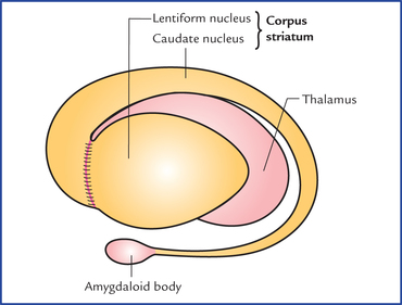
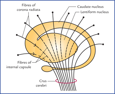
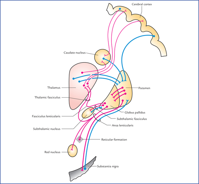
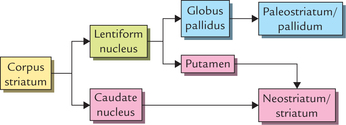
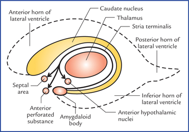
13 The basal ganglia are the large masses of grey matter situated within the white core of each cerebral hemisphere and form essential constituents of the extrapyramidal system. The basal ganglia are now recognized as basal nuclei, but the former term is still commonly used. Anatomically, the term basal ganglia include: The corpus striatum is situated lateral to the thalamus. Topographically it is almost completely divided into the caudate nucleus and the lentiform nucleus by a band of nerve fibres, the internal capsule. However, anteroinferior ends of these nuclei remain connected by a few bands of grey matter across the anterior limb of internal capsule. These bands give it a striated appearance, hence the name corpus striatum (Figs 13.1–13.3). Fig. 13.3 Corpus striatum, thalamus, claustrum and internal capsule as seen in horizontal section of the cerebral hemisphere. The globus pallidus is relatively ancient and termed paleostriatum/pallidum. The caudate nucleus and putamen being recent in development, together form the neostriatum/ striatum. The striatum is largely afferent whereas pallidum is largely efferent structure. These features of corpus striatum may be summarized as follows: Caudate nucleus is a large comma-shaped mass of grey matter, which surrounds the thalamus and is itself surrounded by the lateral ventricle (Fig. 13.4). Its whole length of convexity projects into the cavity of lateral ventricle. Fig. 13.4 Relationship of caudate nucleus with the cavity of the fourth ventricle and thalamus. Note that the stria terminalis, the main efferent tract of amygdaloid body projects to the septal area, anterior perforated substance, and anterior hypothalamus. Its rounded anterior part in front of interventricular foramen is called its head. The head gradually and imperceptibly tapers caudally into the body and then into a tail which merges at its anterior extremity with an almond-shaped mass of grey matter called amygdaloid body (Fig. 13.2). The head is large and rounded, and forms the floor and lateral wall of the anterior horn of lateral ventricle. The bands of grey matter connect it to the putamen across the anterior limb of internal capsule (Fig. 13.3). It has three surfaces and divides into two parts: • The lateral surface is convex and related to thin sheet of white matter, the external capsule. It is grooved by lateral striate arteries (the central branches of middle cerebral artery). • Medial surface is more convex and related to internal capsule (limbs and genu). In transverse sections, the medial surface is angulated at the genu. • Inferior surface is related to sublentiform part of internal capsule and lies close to the anterior perforated substance. The globus pallidus is smaller medial part. It is lighter in colour and consists of large (motor) cells. It is also known as pallidum as it appears pale in section (pallid = pale). The globus pallidus is further subdivided by an internal medullary lamina of white matter into outer and inner segments (Fig. 13.3). The striatum receives afferents chiefly from cerebral cortex, thalamus and substantia nigra. • Corticostriate fibres arise from a wide area of the ipsilateral cerebral cortex and reach the striatum through both internal and external capsules. • Thalamostriate fibres arise from mediodorsal, intralaminar and midline nuclei of the thalamus. The majority of these fibres end in the caudate nucleus, the remaining pass through the internal capsule to reach the putamen. • Nigrostriate fibres arise from substantia nigra and ascend up to terminate in the corpus striatum, mainly in putamen and caudate nucleus.
Basal Nuclei (Basal Ganglia)
Corpus Striatum

Caudate Nucleus
Lentiform Nucleus
Surfaces
Parts
Connections of corpus striatum (Fig. 13.5)
Afferent connections
![]()
Stay updated, free articles. Join our Telegram channel

Full access? Get Clinical Tree


Basal nuclei (Basal Ganglia)