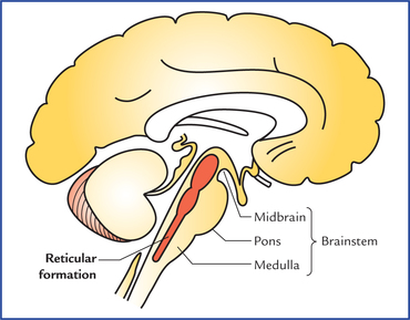
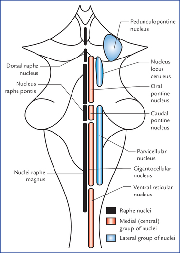
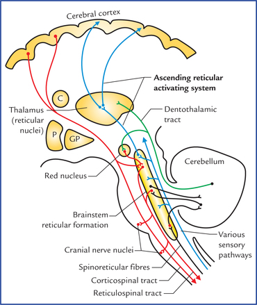
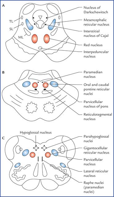
19 The reticular formation is defined as diffuse ill-defined mass of intermingled neurons and nerve fibres occupying the entire core of brainstem (Fig. 19.1). The reticular formation has derived its name from its light microscopic appearance of a vague network of nerve cells and nerve fibres. It has been defined to include all areas within the brainstem (except the named nuclei and tracts) which when stimulated will produce arousal. The knowledge of reticular system is important, because: (a) it regulates levels of consciousness, and alertness, (b) i t regulates respiration, blood pressure, heart rate and other vegetative functions, (c) it regulates tone of skeletal muscles, and The reticular nuclei in brainstem are arranged into three longitudinal columns (Fig. 19.2). • Median column lies in the midline and consists of intermediate size neurons. The nuclei of this column are termed raphe nuclei. • Medial column consists of nuclei which are made up large-size neurons, hence this column is also termed magnocellular column. • Lateral column consists of nuclei which are made up of small neurons, hence this column is also termed parvo-cellular column (parvus = little, small). The nuclei belonging to these columns are shown in Figure 19.2. Since it is not advisable for the student to burden his memory with the names of all the nuclei, only those which have a functional or descriptive value are labelled. The reticular nuclei as seen in transverse sections of the midbrain, pons and medulla are shown in Figure 19.3. Fig. 19.3 The transverse sections of: midbrain (A), pons (B), and medulla (C) showing the location of reticular nuclei. (ML = medial lemniscus, SL = spinal lemniscus, TL = trigeminal lemniscus.) The raphe nuclei form a contiguous column in the mid-line. The neurons of raphe nuclei produce serotonin, a substance that they use as a neurotransmitter. The dorsal raphe nucleus located in the midbrain projects to the spinal cord and forms the pain controlling pathway. The reticular formation receives information from almost all the principal parts of the nervous system and in turn, projects (directly or indirectly) to all these parts (Fig. 19.4). The afferents are classified into three types: – Optic system—through tectoreticular fibres, – Olfactory and limbic systems—through variety of descending pathways, – Auditory system—through tectoreticular fibres, – Spinal pathways—through spinoreticular fibres. A considerable number of fibres of spinothalamic tract terminate in the lateral reticular nucleus of medulla, which in turn project to the cerebellum. Spinoreticulo-cerebellar pathway is an important pathway for carrying exteroceptive sensations to the cerebellum, – Cerebellum from both but mainly from contralateral fastigial nucleus. – Basal ganglia, mainly from corpus striatum. – Thalamus, hypothalamus and subthalamus. – Limbic system, mainly from septal areas, amygdaloid nuclei, and hippocampus. – Cerebral cortex mainly from motor and sensory areas of the cerebral cortex.
Reticular Formation and Limbic System
Reticular Formation
Anatomical Extension
Reticular nuclei in the brainstem
Raphe nuclei (median group of nuclei)
Connections of Reticular Formation
Afferent connections
Afferents from various sensory pathways or systems
Afferent fibres from other parts of central nervous system
Reticular formation and limbic system