CHAPTER 372 Basilar Trunk Aneurysms
Aneurysms of the basilar artery trunk are among the most challenging vascular lesions to treat surgically because of their structural relationship to the brainstem and cranial base. The intricate neurovascular anatomy of the anterior brainstem makes the potential for surgical morbidity relatively high, necessitating the surgeon’s familiarity with surgical approaches to this region. Basilar trunk aneurysms (BTAs) are those located between the vertebrobasilar junction (VBJ) and the take-off of the superior cerebellar arteries (SCAs), frequently arising at the origin of one of many perforating branch vessels of the region. According to a series of nearly 1200 posterior circulation aneurysms treated by Peerless and colleagues, 8% were reported to be BTAs.1
Preoperative consideration of the superior-inferior origin of aneurysms along the basilar artery is imperative and is often characterized in relation to segments of the clivus or expressed as a distance from the dorsum sellae.2 The origin of the anterior inferior cerebellar artery (AICA) is a common site for many of these aneurysms and is therefore also frequently referred to in order to distinguish upper BTAs (those located between AICA and SCA) from lower BTAs (those located between the AICA and VBJ). The origin and size of the aneurysm will in part dictate the surgical approach required to achieve adequate exposure of the aneurysm neck and surrounding neurovascular structures, based primarily on the bony structures that impede the trajectory to the region of interest. Because minimization of brain retraction is a priority in any approach to the basilar trunk, the surgical management of BTAs traditionally employs one of a variety of cranial base approaches with standard microneurosurgical clipping strategies. As with any neurosurgical procedure, the extent of each surgical approach must be weighed against the potential morbidity incurred.
In recent years, endovascular coil embolization has proved to be an increasingly effective and minimally invasive alternative to the surgical clipping of posterior circulation aneurysms and has become the first-line treatment for such lesions at several major institutions.3–5 As a higher proportion of BTAs are successfully coiled by endovascular techniques, however, the subset of aneurysms not amenable to coil embolization that require operative intervention are therefore increasingly complex and challenging to treat from a surgical standpoint. More specifically, this group includes giant, partially thrombosed, and fusiform aneurysms, those with unfavorable neck-to-dome ratios, and those that have failed attempted endovascular coiling.4,6 An ability to perform complex cranial base approaches, in addition to a variety of adjunctive surgical measures that will be discussed, must be at the surgeon’s disposal to safely and efficiently treat these complex aneurysms.
Clinical Presentation
Aneurysms of the posterior circulation frequently present secondary to subarachnoid hemorrhage resulting from aneurysmal rupture. Clinical symptoms such as severe headache, photophobia, nausea and vomiting, and meningismus are typically encountered. More severe clinical sequelae are frequently observed after aneurysm rupture in this region and include syncope, coma, and death.7 The mortality rate associated with ruptured BTAs remains high, nearing 30% in some series.7–11 A variety of cranial nerve palsies and hydrocephalus may additionally be encountered secondary to SAH in the brainstem region.12
Unruptured BTAs frequently present secondary to mass effect on brainstem structures and the related cranial nerves.7 Traditionally, aneurysms of the basilar apex, SCA, and upper basilar trunk may result in oculomotor nerve paresis. Middle BTAs in the region of the AICA may result in abducens nerve paresis, facial nerve paresis, or hearing loss. Those located at the VBJ may present secondary to mass effect on the 9th, 10th, and 11th cranial nerves. Signs of brainstem compression, such as hemiparesis, hemisensory deficits, and gait imbalance, may result from mass effect from unruptured BTAs.11,13 Signs and symptoms of brainstem ischemia or transient ischemic attacks (TIAs) may also be noted.7,14 Finally, BTAs are sometimes discovered incidentally during cranial imaging for a variety of other reasons. As in any patient with an unruptured cerebral aneurysm, the risks of treating the aneurysm are to be weighed carefully against the cumulative risks of conservative management anticipated over the duration of the patient’s life. Our discussion is restricted to saccular lesions. Giant dolichoectasias are considered a separate pathology with nonanalogous natural history and treatment.
Anatomy of Basilar Trunk Aneurysms and Surrounding Structures
The basilar artery originates at the VBJ and courses anterior to the brainstem until it bifurcates in the interpeduncular cistern. The length of the basilar artery varies between 20 and 40 mm, with a mean length of 30 mm.15 Along its course posterior to the clivus, the basilar trunk supplies multiple arteries that are responsible for the perfusion of the brainstem and cerebellum.16 Identification and preservation of the perforating arteries of this region is axiomatic in reducing surgical morbidity when treating BTAs.
The AICA originates several millimeters above the VBJ near the abducens nerve rootlets and courses through the cerebellopontine angle adjacent to the foramen of Luschka to ultimately supply the inferior aspect of the cerebellum, pons, and eighth cranial nerve.15 The SCAs originate within 2.5 mm of the basilar apex and course below the tentorium and adjacent to the superior cerebellar peduncle in the cerebellomesencephalic cistern to supply the superior aspect of the cerebellum.15 Multiple intermediate perforating arteries also arise from the basilar artery, the most proximal being the pontomedullary artery, which arises between the VBJ and AICA.16 Multiple perforating vessels typically arise from the midbasilar trunk, at the level of the pons. These include the superolateral and inferolateral pontine arteries, which supply the paramedian and lateral pontine surface, as well as numerous additional pontine perforators.16 Additionally, several smaller groups of microperforators are found in subgroups known as the caudal, middle, and rostral groups.16 Each group may include up to 10 perforating vessels, with a significant degree of anastomosis within each subtype.
Endovascular Treatment of Basilar Trunk Aneurysms
During the past decade, the ability to treat increasingly complex aneurysms using endovascular coiling procedures has progressed significantly. Some endovascular series have reported excellent outcomes and safety profiles in patients with basilar artery aneurysms.3,5,8,14,17,18 In the largest series to date reporting BTAs treated by endovascular coil embolization, 39 patients had 41 basilar trunk aneurysms treated with a coiling procedure.18 The authors reported excellent or good outcomes in nearly 90% of these patients, with sacrifice of the parent artery in 5 patients.
Complex or fusiform BTAs may not be amenable to clip ligation or direct aneurysm coiling, and may instead require endovascular occlusion of the parent basilar artery.8,19 A balloon occlusion test is performed before complete occlusion, in which multiple modalities are used to demonstrate that the basilar artery can be safely occluded without evidence of brainstem ischemia. These include neuroleptic anesthesia with direct neurological examination, brainstem auditory evoked response (BAER) monitoring, single-photon emission computed tomography (SPECT), and cerebral angiography.8 Furthermore, the size of the posterior communicating arteries is an important prognostic factor in determining a patient’s ability to tolerate complete parent artery occlusion.20
Even more recently, stent-assisted coiling for BTAs has been reported.21 Additionally, the placement of one or multiple porous stents, independent of coiling, has been described as a sole treatment modality for these aneurysms. However, the long-term efficacy of this treatment remains to be determined.
Surgical Treatment of Basilar Trunk Aneurysms
Selection of Surgical Approach to Aneurysms of the Basilar Trunk
Selection of the appropriate surgical approach for a BTA depends on the superior-inferior location of its origin along the basilar artery and its relationship to the clivus and petrous bone. Aneurysms originating from the upper portion of the basilar trunk, frequently at or directly below the bifurcation of the SCA, can be exposed and treated in a manner similar to basilar apex aneurysms. The relationship of the basilar apex to the dorsum sella is a major factor in the preoperative planning of such aneurysms.15 Standard surgical approaches to the upper basilar trunk have included various modifications of the standard transsylvian approach (such as the orbitozygomatic approach), the extradural temporopolar approach, subtemporal transtentorial approach, and anterior transpetrosal approach.2,22–28 Because of the relatively high morbidity rates associated with transpetrosal approaches, these more extensive cranial base procedures have fallen out of favor during the past decade or so. Extended pterional-based approaches, such as the orbitozygomatic (OZ) craniotomy, in conjunction with surgical drilling of the anterior and posterior clinoid processes, is frequently sufficient in achieving adequate exposure of the upper basilar region.11
Aneurysms located in the region of the midbasilar trunk are among the most difficult to approach surgically, because of their location posterior to the midportion of the clivus, distance below the dorsum sellae, and interposed petrous bone. Midbasilar trunk aneurysms are commonly located at the origin of AICA, although less frequently, they may originate from smaller pontine arteries or arise as fusiform lesions. In general, one of several potential transpetrosal approaches is required for the surgical exposure of midbasilar trunk aneurysms. Typical approaches to this region include the combined supratentorial and infratentorial transpetrosal approach, retrolabyrinthine-transsigmoid approach, and anterior transpetrosal (Kawase) approach.2,29–33 Depending on the author, the term transpetrosal implies either removal of varying degrees of the petromastoid bone or ligation and division of the superior petrosal sinus. The retrolabyrinthine, translabyrinthine, and transcochlear approaches are increasingly extensive transpetrosal approaches that can be used in conjunction with various supratentorial trajectories, yet carry higher risks for cranial nerve injury and cerebrospinal fluid (CSF) leakage. In the translabyrinthine and transcochlear exposures, hearing is necessarily sacrificed, whereas the transcochlear approach additionally endangers function of the facial nerve.11
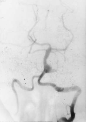
Aneurysms of the lower basilar trunk and VBJ have traditionally been accessed surgically through the retrolabyrinthine-transsigmoid, far-lateral, or extreme lateral inferior transtubercular (ELITE) approaches (Fig. 372-1).4,29–32,34–36 The transoral-transclival approach has also been used to treat aneurysms of this region, yet carries with it greater risks for CSF leakage and meningitis.37,38 In recent years, however, there has been a resurgence of transclival approaches to the anterior brainstem, based on endoscopic exposures with subsequent reconstruction of the skull base using vascularized nasoseptal-based flaps.39–41 Significantly reduced rates of CSF leakage have been achieved using such flaps in the reconstruction of large skull base defects.
Approaches Oriented Parallel to the Basilar Artery
Modifications of the Transsylvian Approach for Basilar Trunk Aneurysms
Various modifications of the original transsylvian approach have been used to treat aneurysms of the upper basilar trunk and basilar apex. Although standard pterional and supraorbital craniotomies can be used to gain access to this region, wider cranial exposures are often required to safely improve visualization of the upper basilar artery region. These modifications have included the addition of an orbitopterional or OZ craniotomy.25,42 The major benefits of these modifications include a widened exposure, redirection of the bulky temporalis muscle, and an improved approach angle that is more parallel to the basilar artery complex. Despite these benefits, the OZ craniotomy is generally limited in reaching basilar artery aneurysms originating below the origin of the SCA and are usually reserved for accessing aneurysms limited to the upper two fifths of the basilar artery.11
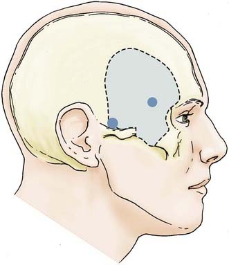
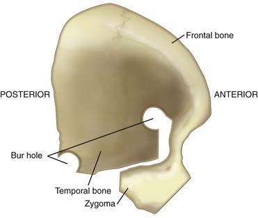
The OZ construct can be removed in a one-piece or two-piece fashion. Osteotomies are required in three locations: (1) at the lateral orbital rim extending to the frontozygomatic suture, (2) at the root of the zygoma parallel to the temporal squama, and (3) at the malar eminence. A frontotemporal craniotomy is performed, with bur holes placed in the frontal and temporal regions (Figs. 372-2 and 372-3). The dura is tacked up to the periphery of the craniotomy, and the sphenoid wing is flattened extradurally using a high-speed drill. The dura is opened in a semilunar fashion and tacked up to the anterior face of the exposure. Standard transsylvian dissection is carried out with the use of the operating microscope, using frontal and temporal retractors as needed for gentle retraction. Additional maneuvers to widen the exposure to the upper basilar region include drilling down the anterior and posterior clinoid processes and dorsum sella as well as retraction of the temporal tip following the coagulation of temporal tip veins. Mobilizing the oculomotor nerve and resecting or retracting the edge of the tentorium further increases the aperture to the upper basilar artery, posterior cerebral arteries, and SCAs.
Extradural Temporopolar Approach
A more expanded approach to the upper basilar artery region is the extradural temporopolar approach. Often used in conjunction with an OZ craniotomy, this approach improves parallel visualization of the basilar artery complex above the origin of the SCA. A significant degree of drilling of the middle skull base is required to untether and retract the temporal and frontal lobes and expose the posterior petroclival region. The major benefit offered by this approach is extradural retraction of the temporal and frontal lobes, which provides a significant margin of brain protection compared with intradural retraction. After initial exposure, a sequential progression of extradural drilling, untethering, and retraction is carried out, including (1) drilling of the sphenoid wing and lateral aspect of the superior orbital fissure (SOF), (2) drilling of the anterior clinoid process, (3) unroofing of the optic canal, (4) mobilization and retraction of the temporal tip, (5) transcavernous mobilization of the internal carotid artery (ICA) and cranial nerves, and (6) drilling of the posterior clinoid process.22,23
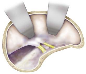
Starting with a standard OZ craniotomy, as previously described, the temporal lobe is elevated extradurally to expose the SOF and foramen rotundum (Fig. 372-4). The limitations of dural retraction at this point include the superior ethmoidal artery and foramen ovale. The sphenoid wing is drilled down, followed by drilling of the lateral wall of the SOF, which is skeletonized to gain access to the junction of the dural sleeve and the dura propria of the temporal lobe. The foramen rotundum and optic canal are unroofed for the same reason. The anterior clinoid process is drilled down extradurally. The temporal tip is retracted to expose the junction of the dura propria of the temporal lobe and the true inner cavernous membrane. The meningo-orbital vessels are coagulated and divided. This dural plane is then carefully developed with special attention directed to preserving the sheaths over the third, fourth, and fifth cranial nerves. As the outer layer of the cavernous sinus is dissected, hemostasis is maintained with gentle packing of Surgicel.


Stay updated, free articles. Join our Telegram channel

Full access? Get Clinical Tree


