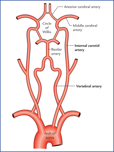
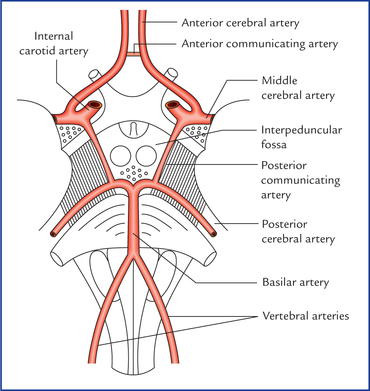
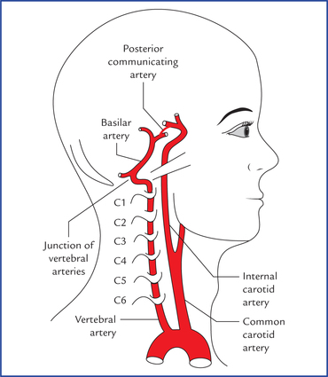
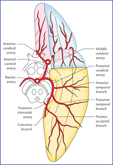
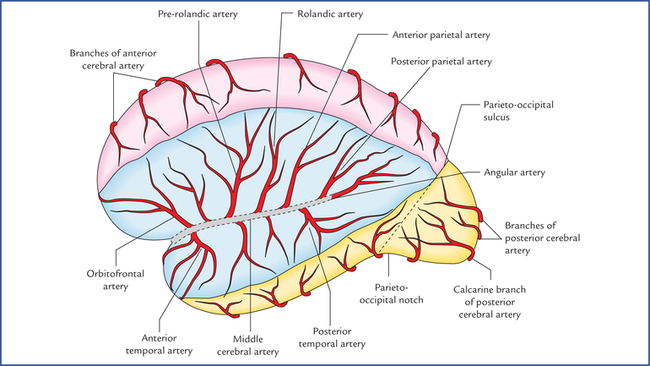
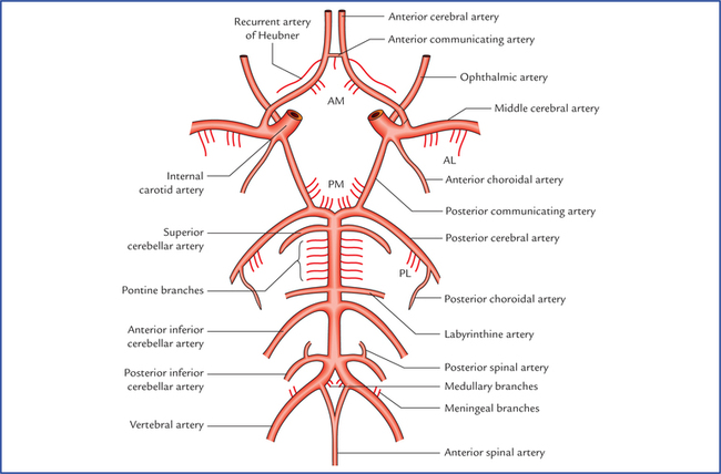
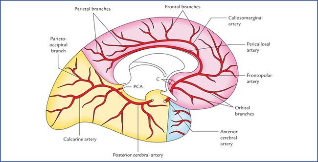
15 The continuous blood supply to the brain is of utmost importance because of its high metabolic demands for oxygen and glucose. It is highly sensitive to hypoxia (inadequate O2) and hypoglycaemia (subnormal concentration of glucose in the blood). The consciousness is lost within 10 seconds of cessation of blood flow, and if the state continues, an irreversible brain damage starts to occur at about 4 minutes and is completed within 10 minutes. The brain is supplied by the paired internal carotid and vertebral arteries via an extensive system of branches (Fig. 15.1): The major arteries supplying the cerebrum (i.e. branches of basilar and internal carotid arteries) get interconnected to one another at the base of the brain to form a six-sided polygon of arteries called circulus arteriosus or circle of Willis (Figs. 15.2 and 15.3). The circle of Willis is formed around the interpeduncular fossa and lies in the interpeduncular subarachnoid cistern. It contributes most of the arterial blood supply to the brain. Fig. 15.3 Circle of Willis and the branches of arteries supplying the brain. The central branches of cerebral arteries are shown by abbreviations: AM = anteromedial group, PM = posteromedial group, AL = anterolateral group, PL = posterolateral group. Anteriorly, by the anterior communicating and the anterior cerebral arteries. Posteriorly, by the basilar artery dividing into two posterior cerebral arteries. Laterally on each side, by the posterior communicating artery connecting the internal carotid artery with the posterior cerebral artery. Normally there is little or no mixing of blood streams: (a) of two vertebral arteries in the basilar artery, (b) of two anterior cerebral arteries in the anterior communicating artery, and (c) of internal carotid and posterior cerebral arteries in the posterior communicating artery. Therefore, right half of the brain is supplied by right vertebral and right internal carotid arteries and left half of the brain is supplied by left vertebral and left internal carotid arteries. The vertebral artery, a branch of subclavian artery, ascends in the foramina transversaria of upper six cervical vertebrae. On reaching the base of skull, it winds backwards and medially around the lateral mass of the atlas and pierces posterior atlanto-occipital membrane, to enter the posterior cranial fossa through the foramen magnum where it runs on the anterolateral aspect of the medulla. Here the two vertebral arteries converge, and unite at the lower border of the pons to form the basilar artery (Fig. 15.4). • Anterior spinal artery is a small branch arising near the termination of the vertebral artery. It descends in front of the medulla and unites with its fellow of the opposite side at the level of the lower end of the olive to form a single median trunk that descends along the anterior longitudinal fissure of the spinal cord. • Posterior spinal artery arises from vertebral artery and sometimes from posterior inferior cerebellar artery. It passes downwards on the posterior surface of the spinal cord, after dividing into two branches; one along the medial side, and the other along the lateral side of the dorsal roots of the spinal nerves. • Posterior inferior cerebellar artery is the largest branch of the cranial (4th) part of the vertebral artery. It arises near the lower end of the olive, winds backwards around the medulla oblongata, and then ascends to the pon-tomedullary junction. • Meningeal branches are small and supply the dura mater of the posterior cranial fossa. • Medullary arteries are several minute vessels which supply the medulla oblongata. • Pontine branches are numerous short slender parame-dian vessels which pierce the pons to supply it. • Anterior inferior cerebellar artery arises close to the lower border of the pons and runs backwards and laterally usually ventral to the VIIth and VIIIth cranial nerves. Then it forms a loop over the flocculus of the cerebellum and peeps into the internal acoustic meatus for a variable distance lying below the VIIth and VIIIth cranial nerves. After exit from the meatus it supplies the anterolateral portion of the inferior surface of the cerebellum. • Labyrinthine artery is a long slender branch which arises either from basilar artery or from anterior inferior cere-bellar artery. It accompanies the vestibulocochlear nerve and enters the internal auditory meatus to supply the internal ear. It is an end artery. • Superior cerebellar artery arises close to the superior border of the pons, runs laterally below the oculomotor nerve (which is interposed between this artery and the posterior cerebral artery), and winds round the cerebral peduncle below the trochlear nerve to reach the superior surface of the cerebellum which it supplies. • Posterior cerebral artery passes laterally parallel to the superior cerebellar artery, curves around the midbrain to reach the medial surface of the cerebral hemisphere, beneath the splenium of corpus callosum (Figs 15.5 and 15.6). The artery gives off temporal branches which ramify over the inferior surface of the temporal lobe, and calcarine and parieto-occipital branches which run along the corresponding sulci. Fig. 15.6 Arteries on the inferomedial aspect of the cerebral hemisphere. (C = central branches, PCA = posterior choroidal artery.) The internal carotid artery, a terminal branch of the common carotid artery, traverses the carotid canal in the base of the skull and enters the middle cranial fossa beside the dorsum sellae of the sphenoid bone. Here it first runs forwards along the floor and medial wall of the cavernous sinus and then turns upwards on the medial side of the anterior clinoid process. At this point the artery pierces the dural roof of the cavernous sinus and also the arachnoid mater to enter the subarachnoid space. Now it first runs backwards and then upwards to come to lie lateral to the optic chiasma just underneath the anterior perforated substance of the brain, where it terminates by dividing into two branches, a larger middle cerebral artery and a smaller anterior cerebral artery (Figs 15.2 and 15.3). • Ophthalmic artery arises from the ventral convexity of the carotid siphon and enters the optic canal to reach the orbital cavity to supply the structures of the orbit including eyeball. • Posterior communicating artery arises close to the termination of the internal carotid artery. It runs backwards and anastomoses with the proximal part of the posterior cerebral artery. • Anterior choroidal artery is a long slender branch, which arises just distal to the origin of the posterior communicating artery. It courses backwards above and along the optic tract, to enter the inferior horn of the lateral ventricle through the choroid fissure to end in the choroid plexus. • Anterior cerebral artery is a smaller terminal branch of the internal carotid artery. It runs forwards and medially above the optic nerve to the commencement of the median longitudinal cerebral fissure, where it comes very close to its fellow of the opposite side and gets joined with it by a short transverse anterior communicating artery. The anterior cerebral artery then curves around the genu of corpus callosum. • Middle cerebral artery is the larger terminal branch of the internal carotid artery. It appears to be the direct continuation of the internal carotid artery and carries about 30% of the carotid blood flow. The middle cerebral artery first runs laterally in the stem of the lateral sulcus (Fig. 15.5) and then turns backwards and upwards in the posterior ramus of the lateral sulcus, where it breaks up into frontal, parietal and temporal branches which emerge from the lateral sulcus and run towards the areas of their supply. Some of these branches are named (Fig. 15.7). The branches of main arteries supplying the brain are summarized in Table 15.1 Table 15.1 Branches of the main arteries of the brain
Blood Supply of the Brain
Arteries of The Brain
Circle of Willis (circulus arteriosus)
Functional significance of circle of Willis
Vertebral System
Vertebral artery (Fig. 15.4)
Branches of the cranial part of the vertebral artery (Fig. 15.3)
Basilar artery (Figs 15.1–15.3)
Branches of basilar artery
Carotid System
Internal carotid artery (Fig. 15.4)
Branches of the cerebral part of the internal carotid artery (Fig. 15.3)
Cerebral part of internal carotid artery
Fourth part of vertebral artery
Basilar artery
• Ophthalmic artery
• Meningeal arteries
• Anterior inferior cerebellar artery
• Anterior cerebral artery
• Anterior spinal artery
• Labyrinthine artery
• Middle cerebral artery
• Posterior spinal artery
• Pontine arteries
• Posterior communicating artery
• Posterior inferior cerebellar artery
• Superior cerebellar artery
• Anterior choroidal artery
• Medullary arteries
• Posterior cerebral artery ![]()
Stay updated, free articles. Join our Telegram channel

Full access? Get Clinical Tree