Fig. 7.1
Diagrammatic example of the non-injective nature of the relation between cognitive states and neuroimaging (fMRI) activation maps – given the current spatial resolution of the technique
7.4 Cogito Ergo Sum by fMRI1
Determining the degree of residual cognitive processing that may be available in patients who survive severe BI is very important and can assist, in the rehabilitative context, in determining which modalities might be – at least potentially – available to try to elicit responses in a patient and, where voluntary responses are detected, to harness them into methods of communication. An even more pressing – and more controversial – question, however, is whether neuroimaging responses can be taken to weigh on the determination of whether a patient is (at least minimally) conscious. In the insightful words of A.H. Ropper [24], is “cogito ergo sum by fMRI” possible (and/or admissible in the clinical context; see Chap. 12)? While the answer to this question is tied to several of the complexities mentioned above, many agree that one of the most important variables in adjudicating the issue of whether neuroimaging activations can index the presence of awareness is the nature of the experimental design employed (see [23, 25]). On the one hand, detecting different patterns of brain activation in response to different sensory stimulations, as in the picture of a face versus the picture of a house (see Fig. 7.2, top row), might be taken to imply that a brain possesses sufficient bottom-up mechanisms to distinguish the two stimuli (with no implication as to whether the patient has any subjective experience relating to them).
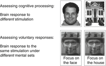
Fig. 7.2
Top row: Comparison of brain response to different sensory stimulations only allows inferring, generally speaking, “appropriate” bottom-up residual processing. Bottom row: Comparison of brain responses to the same sensory stimulation, but under different mental sets, allows inferring the presence of voluntary (i.e., top-down) responses (Adapted from [7])
On the other hand, detecting different patterns of brain activation in response to the same stimulus, when under different instructions (i.e., mental sets), can only be explained – once sources of artifactual activation such as motion are excluded – by someone voluntarily complying with the instructions and engaging in top-down voluntary cognitive processes, both of which imply a state of (at least minimal) awareness. To exemplify, consider the ambiguous stimulus depicted in the bottom row of Fig. 7.2. If a patient consistently demonstrates, in response to the same ambiguous picture, sustained (i.e., 16 s) upregulation of the face selective area in the fusiform gyrus and contemporaneous downregulation of the place-sensitive parahippocampal area in the periods in which she/he is asked to focus on the face in the image, and the reverse pattern in the periods in which she/he is asked to focus on the house in the image,2 the simplest explanation is that the patient has understood the instructions and is performing the expected task.
In a landmark paper, in 2006, this logic was employed to demonstrate, for the very first time, that it is possible for a patient to appear unresponsive during bedside clinical assessments while being able to engage in voluntary top-down mental activities (e.g., “imagining playing tennis”), as captured by fMRI [26]. Specifically, a patient exhibited appropriate and sustained (for 30 s) brain activations, matching those seen in healthy volunteers, in response to short single-word cues (e.g., “tennis”) when, by all clinical criteria, no sign of voluntary responsiveness or awareness could be detected. Since this pioneering paper, a number of publications have replicated the result in the context of similar experimental designs [27, 28], different experimental designs [29, 30], as well as different methodologies [31, 32].3 While groundbreaking at the time, this finding is not necessarily unexpected. A number of previous studies, including retrospective audits [33, 34] as well as comparative evaluation of the diagnostic accuracy of different patient assessment techniques [35, 36], had already shown that misdiagnosis – by which (minimally) conscious patients are diagnosed as vegetative – is more frequent than desirable. While the relative rarity of these conditions, inconsistent terminology, and lack of specialized training were recognized early on as important causes of misdiagnosis [34], it is now also understood that the diagnosis of VS rests on a logical flaw [22]: absence of observable evidence of awareness (i.e., failure of a patient to demonstrate any recognizably voluntary behavior) cannot be taken to necessarily imply absence of awareness. For, a patient could be (minimally) conscious, but unable to respond because of sensory or motor impairments or transient unconsciousness, or even just unwillingness to respond [4].
7.5 Important Caveats
As the potential of neuroimaging to uncover signs of awareness in otherwise behaviorally unresponsive patients is more and more recognized, it is important to be mindful of a number of important issues concerning the interpretation of neuroimaging data in this context.
7.6 Positives and Negatives: Dissociation Is a 2-Way Street
As briefly reviewed above, a number of studies have shown that it is possible for a patient to appear unresponsive in (behavior-based) bedside clinical testing while being responsive in neuroimaging assessments [26, 27, 29, 30, 32]. A number of recent studies, however, have reported instances of the reverse dissociation: some patients can demonstrate a state of consciousness at the bedside but fail to show any significant activation during “active” neuroimaging sessions. Bardin and colleagues, for example, have shown that only half of a group of (at least minimally) conscious patients, as determined by clinical testing, could demonstrate significant activity in mental imagery tasks [37]. Similarly, in a recent study of top-down processes in DOC patients, while 3 out of 8 patients with a VS diagnosis could demonstrate voluntary (brain) behavior during neuroimaging sessions, only 6 out of 12 MCS patients4 and 3 out of 4 exit-MCS patients could not demonstrate any significant activation [29]. In a large cohort study, the overall sensitivity of the “imagery” task in fMRI to detect a state of MCS was recently estimated at about 45 % [28]. The existence of two-directional dissociations highlights two important aspects of the use of neuroimaging in the context of DOC. On the one hand, it confirms that there are instances in which neuroimaging can uncover voluntary brain responses in a subset of patients who appear behaviorally (i.e., at the bedside) unresponsive. On the other hand, these dissociations imply that false negatives are as possible in neuroimaging assessments as they are in behavioral assessments (although it is currently unknown how the two rates compare). In other words, negative neuroimaging results should not be interpreted.
7.7 The “Tip of the Iceberg” Problem
An important, but not very appreciated, issue, in the use of neuroimaging to assess DOC patients, is the “tip of the iceberg” phenomenon. The kind of “active” tasks (e.g., mental imagery [26], target monitoring [38]) currently employed to covertly detect the presence of consciousness with neuroimaging requires the concurrent presence of a number of cognitive processes in addition to a state of awareness. To name a few, patients must, at a minimum, retain language capabilities sufficient to comprehend a set of instructions; memory functions sufficient to allow maintaining a set of instructions throughout an experimental run; sensory resources, in all the relevant modalities, sufficient to allow processing stimuli; as well as executive functions sufficient to allow, for example, periodic engaging and disengaging in the relevant mental task. Although many of these problems are common to standard clinical assessments, they further stress the importance of not interpreting negative results as evidence of unconsciousness, as well as the need to develop non-language-based and, ideally, “passive” neuroimaging tests capable of detecting neural markers of a conscious state (e.g., [15, 39, 40]).
7.8 Unconscious or in a “Living Hell”: A (Probably) False Dichotomy
Finally, a last important caveat applies to the over-interpretation of evidence (neuroimaging or otherwise) in the context of DOC. In the 1990 ruling of the Supreme Court of the United States (497 US 261) in the case of Cruzan v. Director, Missouri Department of Health, Judge Blackmar, after pointing out that the patient in question, Nancy Cruzan, might have exhibited (probably reflexive) responses to noxious stimulation, noted that “If she has any awareness of her surroundings, her life must be a living hell.” While we will not visit the important legal and ethical issues surrounding DOC (see Chap. 14), there is an important neuroscientific consideration to make with respect to how much mental life should be attributed, in the absence of direct evidence, to patients demonstrating neuroimaging (or for that matter, behavioral) responses. Often, because most fMRI experiments benchmark the activations seen in patients to those observed in healthy volunteers (e.g., [7, 26, 29, 37]), it is tempting to infer that, where matching activations are seen, these imply that the patient might possess the same state of awareness of healthy individuals. While it is certainly not impossible for a behaviorally unresponsive patient to retain normal consciousness (as would be in the very different condition of complete locked-in syndrome), it must be recognized that the brain of an MCS patients, even if capable of supporting some level of consciousness, is nonetheless structurally and functionally severely pathological and extremely different from that of a healthy individual. PET data, for example, have clearly shown that the MCS brain is severely hypometabolic, presenting a cerebral metabolic rate of glucose at about 55 % that observed in healthy volunteers. In fact, although local differences between conscious and unconscious patients can be detected, MCS metabolic rate is much closer to that of a VS patients (estimated at approximately 42 % that of the healthy brain) than fully conscious, healthy, individuals [41, 42] (see Fig. 7.3).
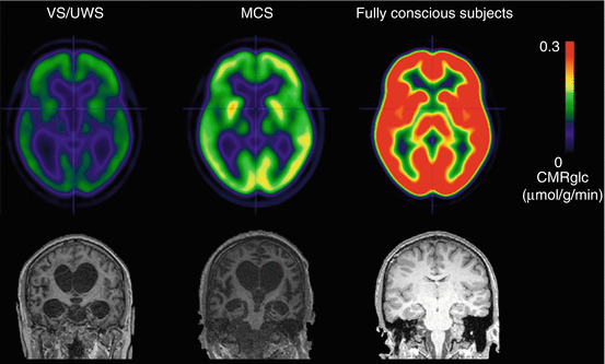
Fig. 7.3
Top: Depiction of average metabolic rates observed in VS, MCS, and healthy volunteers (Adapted from [42] under Creative Commons CC-BY license). Bottom: Sample T1-weighted structural images for a VS, MCS, and healthy volunteer, viewed in a coronal cut (note: the brains shown in the bottom row do not correspond to the brains in the top row and are only meant to represent a sample individual from each group)
Similarly, as illustrated in the bottom row of Fig. 7.3, despite being (at least minimally) conscious, MCS patients present severe widespread pathology as compared to healthy volunteers, which has been measured in terms of both subcortical atrophy [43] as well as degradation of cortico-cortical and cortico-subcortical connectivity [44]. Thus, observing in a DOC patient, voluntary brain responses matching those seen in healthy volunteers should not be automatically taken, by itself, to imply an equivalence between the state of consciousness of the patient and that of a healthy individual – which should, however, not be excluded a priori [45]. Rather, the full power of neuroimaging techniques could be employed, in otherwise nonresponsive patients, to address in an evidence-based way the question of which cognitive resources a patient might retain [46].
7.9 Conclusion
In all, regardless of where one falls on the many issues raised above, there is little doubt that the use of in vivo neuroimaging has greatly benefited the field of disorders of consciousness. First, in the past 18 years, these techniques have started revealing the (at first surprising) extent to which cortical processing can be retained in VS as well as MCS patients despite having sustained a catastrophic brain injury (e.g., [7–9, 11–14, 47–50]). These results fully highlight the ramifications, in the clinical context, of our limited scientific understanding of what consciousness is and our inability to directly measure it or quantify it objectively [22, 23]. Furthermore, neuroimaging has also shown that it is possible for a patient to appear, clinically (i.e., behaviorally), unresponsive while being in fact (at least) minimally conscious, as indexed by the ability to voluntarily engage and disengage, in response to verbal commands, in top-down mental tasks (e.g., [26, 27, 29, 30, 32]). It is crucial to stress that these (to date relatively few) cases in which a dissociation has been reported between the level of awareness observable at the bedside and that observable in neuroimaging assessments do not only reflect the natural challenges tied to assessing patients that have sustained severe brain injuries [33–36]. Indeed, as discussed above and elsewhere [4, 23, 25], no matter how skilled an assessor, a (minimally) conscious patient unable to manifest, through muscle-dependent responses, her state of awareness – due, for example, to motor impairment – would be impossible to distinguish from a VS patient on the basis of clinical protocols. It is in these cases that the full value of neuroimaging becomes evident.
Still, it is also undeniable that the use of neuroimaging techniques in the context of disorders of consciousness requires careful consideration of a number of issues. First, the interpretation of brain activations is necessarily secondary to the specific experimental paradigm employed [25]. Only under certain experimental circumstances (unless convergent evidence from other methodologies, such as anesthesia, is available; see [20]) can brain responses be taken to mark the presence of a state of minimal awareness. Second, only positive evidence should be interpreted, because negative findings are unable to differentiate cases of truly VS patients from cases of MCS patients unable (or unwilling) to respond. Finally, while DOC patients exhibit highly pathological brain function and structure [42, 43, 51], it is also true that the human brain can maintain surprisingly high level of functioning even in the presence of severely pathological features (e.g., [45]; see also the degree of pathology evident in some “responsive” DOC patients [27, 29, 30, 37]). Thus, wherever behavior-based evidence cannot be obtained, neuroimaging might be the one approach capable of assessing which, and how many, cognitive processes can be imputed to any given DOC patient [46].
References
1.
Monti MM, Laureys S, Owen AM (2010) The vegetative state. BMJ 341:c3765. doi:10.1136/bmj.c3765 CrossRefPubMed
Related posts:
 Responsiveness in DoC: A Quest for Consciousness?
Clinical Evaluation of Residual Brain Function and Responsiveness in Disorders of Consciousness
Moving Beyond End of Life: The Ethics of Disorders of Consciousness in an Age of Discovery and Uncertainty
Advances in the Scientific Investigation of Consciousness
Decoding Thoughts in Disorders of Consciousness
Mesocircuit Mechanisms Underlying Recovery of Consciousness Following Severe Brain Injuries: Model and Predictions
Responsiveness in DoC: A Quest for Consciousness?
Clinical Evaluation of Residual Brain Function and Responsiveness in Disorders of Consciousness
Moving Beyond End of Life: The Ethics of Disorders of Consciousness in an Age of Discovery and Uncertainty
Advances in the Scientific Investigation of Consciousness
Decoding Thoughts in Disorders of Consciousness
Mesocircuit Mechanisms Underlying Recovery of Consciousness Following Severe Brain Injuries: Model and Predictions
Stay updated, free articles. Join our Telegram channel

Full access? Get Clinical Tree


