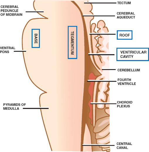
6 Brainstem The brainstem comprises the medulla, pons, and mid-brain. Like the spinal cord, with which it is caudally contiguous, it contains multiple long ascending and descending pathways that are oriented parallel to the long axis of the brainstem, as well as nuclei whose axons are oriented parallel to the transverse plane. There are four major elements of the brainstem: (1) long ascending and descending tracts, (2) cranial nerve nuclei and their fascicles, (3) cerebellar nuclei and their connections, and (4) reticular neurons and their processes. Because brainstem dysfunction is both common and potentially life threatening, prompt recognition of its cardinal manifestations is imperative. An understanding of the principles of the brainstem’s organization simplifies the study of its complex regional anatomy and offers the clinician a logical approach to anatomic diagnosis. This chapter outlines the basic organization of the brainstem, then discusses the regional and functional anatomy of the cranial nerve nuclei, the long ascending and descending tracts, the cerebellar connections, and the reticular formation. A description of the blood supply of the brainstem is followed by a discussion of the localization of brainstem lesions, and finally by a description of classic brainstem syndromes. Grossly, four major parts of the brainstem are contiguous throughout the length of the medulla, pons, and mid-brain (Fig. 6.1): Fig. 6.1 Brainstem. Ten of the 12 cranial nerves have their nuclei in the brain-stem. (Cranial nerves I and II are the exceptions.) Like the spinal gray nuclei, the cranial nerve nuclei are grouped into longitudinal columns. These columns are both anatomically and functionally distinct: medial columns contain exclusively motor nuclei, and lateral columns contain exclusively sensory nuclei. This organization is explained by developmental events, as follows. The alar and basal plates of the developing neural tube give rise to sensory afferents and motor efferents, respectively. Early in development, these plates are positioned in a dorsal/ventral orientation, an orientation that is maintained in the mature spinal cord. In the developing brainstem, however, this organization is changed: the lateral spread of the fourth ventricle causes the dorsal alar plate to rotate laterally in relation to the ventral basal plate. As a result, the lateral columns of the mature brainstem contain strictly sensory cranial nerve nuclei; the medial columns contain strictly motor cranial nerve nuclei. The medial columns are composed of somatic and visceral motor nuclei. Two types of visceral motor nuclei are distinguished on the basis of the two types of muscles their neurons innervatestriated muscle derived from primitive branchial arches and smooth muscle associated with viscera and glands. Three types of motor nucleione somatic, two visceralthus comprise the three medial columns. The individual nuclei that form these columns, in medial to lateral order, are described in the following sections. See Fig. 6.2. This column, which is not continuous longitudinally, is immediately adjacent to the midline, just below the floor of the ventricular system. It contains nuclei composed of neurons that innervate the striated muscles of the head and neck derived from embryonic myotomes (i.e., the extraocular muscles and the muscles of the tongue). In rostrocaudal order, the nuclei contained in this column include the following four structures: Fig. 6.2 Somatic motor nuclei. See Fig. 6.3. This column is located lateral and ventral to column 1. It contains nuclei composed of neurons that innervate the striated muscles of the head and neck derived from the branchial arches (i.e., the muscles of mastication, the muscles of facial expression, the muscles of the pharynx and larynx, and the sternocleidomastoid and trapezius muscles). In rostrocaudal order, the nuclei in this column include the following four structures: Fig. 6.3 Visceral motor nuclei (striated muscle). See Fig. 6.4. This column is located immediately lateral to column 1. It contains nuclei of preganglionic parasympathetic neurons that innervate the smooth muscles and glands of the head and neck, as well as the thoracic and parts of the abdominal viscera. (Preganglionic parasympathetics originating in sacral segments of the spinal cord supply the rest of the abdominal and pelvic viscera.) In rostrocaudal order, these parasympathetic nuclei include the following three structures: Fig. 6.4 Visceral motor nuclei (smooth muscle). The lateral columns are composed of sensory nuclei. Unlike most motor nuclei, whose axons are carried in a single corresponding cranial nerve, each sensory nucleus receives input from several different cranial nerves. There are three major sensory nuclei in the brainstem (Fig. 6.5): Fig. 6.5 Sensory nuclei. Four long tracts, two ascending, two descending, provide landmarks along the transverse axis of the brainstem. The spinothalamic tract lies laterally in the brainstem, whereas the medial lemniscus, the corticospinal tract, and the corticobulbar tract lie medially. See Fig. 6.6. The lateral and anterior spinothalamic tracts are responsible for pain, temperature, and light touch sensation. They are located in the lateral aspect of the teg-mentum throughout the brainstem, adjacent to the descending sympathetic tract. They occur in essentially the same position they occupy in the spinal cord. The spinothalamic tract consists of second-order neurons that originate in the dorsal gray horn of the spinal cord, cross the midline in the anterior white commissure, and project to the ventral posterolateral (VPL) nucleus of the thalamus. Third-order neurons in the VPL thalamus send axons to the postcentral gyrus. Because of the close proximity of the spinothalamic tract to the descending sympathetic fibers, both systems are typically impaired as a result of damage to the lateral tegmentum, where they represent important landmarks. An ipsilateral Horner syndrome (descending sympathetic lesion) is thus often associated with a contralateral hemisensory loss (spinothalamic lesion), which may be caused by a lesion in the lateral medulla or pons. Fig. 6.6 Spinothalamic tract. See Fig. 6.7. The medial lemniscus, which is the rostral continuation of the dorsal columns of the spinal cord, mediates position sense and discriminative touch. It consists of second-order neurons that originate in the nucleus cuneatus and nucleus gracilis. These nuclei receive input from the spinal cord via the cuneate and gracile fasciculi (dorsal columns), which carry impulses from the upper and lower extremities, respectively. After synapse in the ipsilateral cuneate and gracile nuclei, these axons act as the internal arcuate fibers and ascend to the contralateral VPL thalamus. From here they ascend to the sensory cortex. The medial lemniscus is situated in the medulla close to the midline between the posteriorly situated medial longitudinal fasciculus (MLF) and the anteriorly situated corticospinal and corticopontine tracts. In its rostral ascent, the medial lemniscus moves laterally but remains an important landmark of the medial aspect of the medulla and pons.
The Brainstem Is Organized into Four Major Parts
Cranial Nerve Nuclei Are Organized into Longitudinal Columns
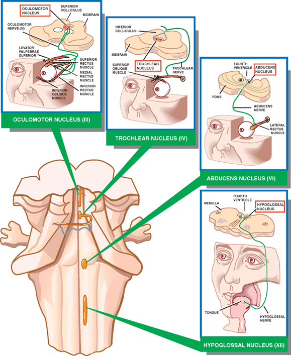
Medial Columns Contain Three Types of Motor Nuclei
Column 1
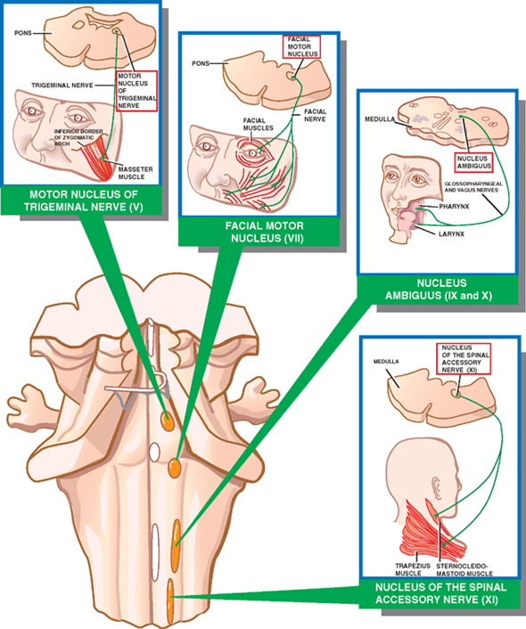
Column 2
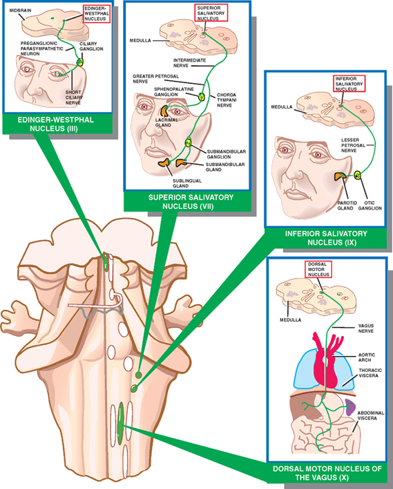
Column 3
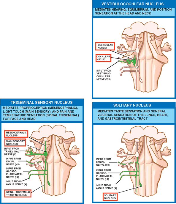
Lateral Columns Contain Three Sensory Nuclei
Long Ascending and Descending Tracts Traverse the Brainstem
Two Ascending Tracts Occur Laterally and Medially
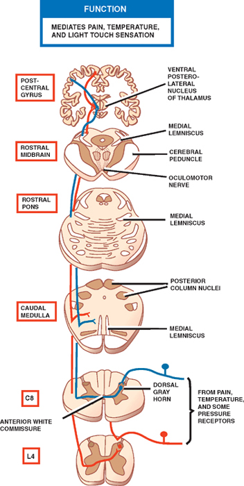
Spinothalamic Tract
Medial Lemniscus

Related posts:


Stay updated, free articles. Join our Telegram channel

Full access? Get Clinical Tree


