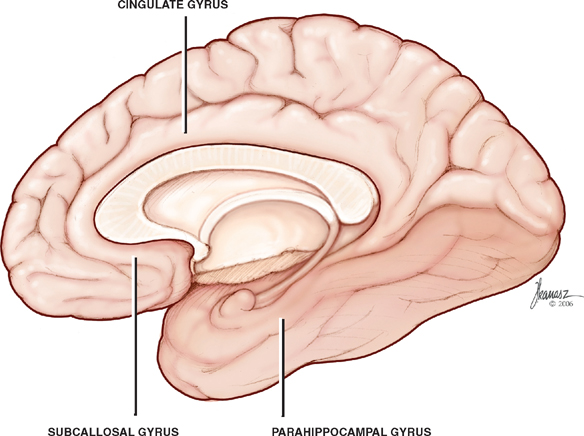
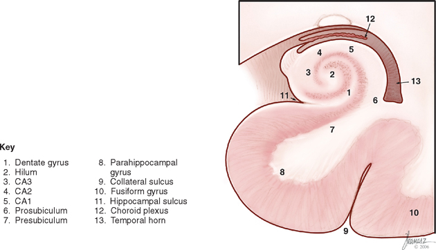
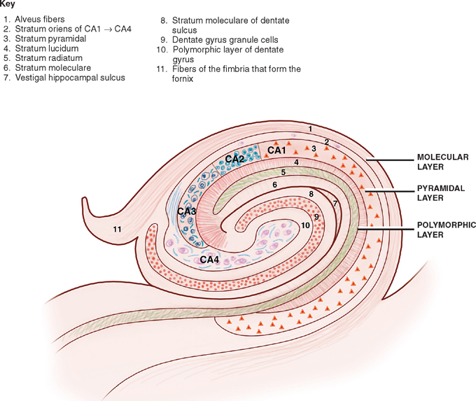
12 Limbic System Many structures have been included in various accounts of the limbic system, but for our purpose the limbic system means (1) the limbic lobe, (2) the hippocampal formation, (3) the amygdaloid nucleus, (4) the hypothalamus, and (5) the anterior nucleus of the thalamus. Functional studies suggest the limbic system participates in emotion and emotional behaviors (e.g., anger, fear, and sexuality), which appear to be important in the preservation of the individual and of the species. In addition, these studies suggest the limbic system is important in coordinating the emotion and autonomic responses such as changes in blood pressure, respiration, pupillary dilation, and bowel and bladder control. More recently appreciated is the important role of the limbic system, in particular the hippocampus, regarding learning and memory. It should be emphasized that, despite the persistent use of the term limbic system, which includes, as noted, several structures that appear to share some common functions, the idea that it represents an independent functional system can no longer be maintained. Indeed, the individual structures of the limbic system are now known to be involved in a wide variety of functions and connections. With this in mind, the major components of the lim-bic system are addressed separately. Further, because the thalamus and the hypothalamus are discussed at length elsewhere (Chapters 9 and 10), this chapter concentrates on the limbic lobe, the hippocampal formation, and the amygdaloid nucleus. In 1878, the French anthropologist Pierre Paul Broca introduced the term limbic lobe to describe a ring of gray matter that surrounded the rostral brainstem. The term, which comprised the subcallosal, the cingulate, and the parahippocampal gyri, was applied to characterize these structures as phylogenetically primitive. In 1937, the American anatomist James Papez transformed the limbic lobe from an evolutionary concept into a physiological one. In an attempt to explain the relationship between consciousness and the emotions, he described a circuit of neuronal connections between the limbic lobe and the hypothalamus. The circuit of Papez is described later in this chapter. One of the historical consequences of Papez’s efforts was to enlarge the concept of a “limbic lobe” into a “limbic system,” which encompasses the limbic lobe as well as associated subcortical structures. See Fig. 12.1. As noted, the limbic lobe consists of (1) the subcallosal, (2) the cingulate, and (3) the parahippocampal gyri. Collectively, these gyri form a ring around the rostral portion of the brainstem. The limbic system is formed by the limbic lobe and associated subcortical structures, the amygdala, habenula, mammillary bodies, septal nuclei, portions of the thalamus, hypothalamus, and midbrain. The hippocampus is part of the limbic lobe and is situated on the inferomedial aspect of the hemisphere in the temporal lobe. Fig. 12.1 Limbic lobe. See Figs. 12.2 and 12.3. The hippocampal formation consists of (1) the hippocampus, (2) the dentate gyrus, and (3) the parahippocam-pal gyrus. The hippocampus has the appearance of a sea horse. It bulges out into the temporal horn of the lateral ventricle and is arched around the mesencephalon. It is divided into three anatomic segments: head, body, and tail. Fig. 12.2 The hippocampus. Fig. 12.3 Hippocampal micro-anatomy. The hippocampus is a bilaminar archicortical (archi-cortex) structure consisting of Ammon’s horn (hippocampus proper) and the dentate gyrus with one lamina rolled up in the other. The hippocampus is composed of an intraventricular expansion of the temporal lobe cortex. It forms the floor of the inferior horn of the lateral ventricle and is C-shaped in the coronal section. Anteriorly, it enlarges to form the pes hippocampus. Posteriorly, it terminates beneath the splenium of the corpus callosum. Histologically, the hippocampus is an example of the three-layered archicortex, which is phylogenetically older than the six-layered neocortex (Fig. 12.4). The three layers of the hippocampus are described as follows: Fig. 12.4 Hippocampal histology. See Figs. 12.5, 12.6, 12.7, and 12.8. Afferent fibers are received from the entorhinal area (lateral olfactory cortex), the septal area, the anterior thalamic nucleus, and the mammillary bodies. Noradren-ergic fibers are projected from the locus ceruleus, and serotonergic fibers are projected from the raphe nuclei. Finally, the fornix carries commissural fibers that originate in the hippocampus on the opposite side. The predominant efferent fibers of the hippocampus are projected in the fornix as follows:
Structures of the Limbic System
Limbic Lobe
Hippocampal Formation
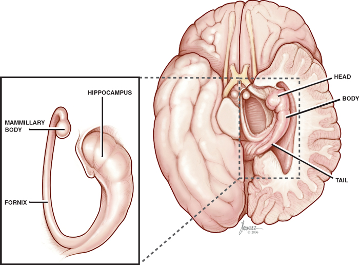
Hippocampus
Histology of the Hippocampus
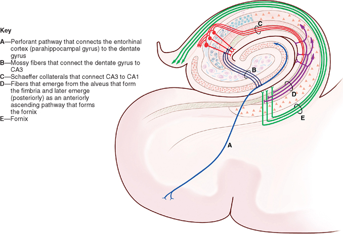
Afferent and Efferent Connections
Related posts:

Stay updated, free articles. Join our Telegram channel

Full access? Get Clinical Tree


