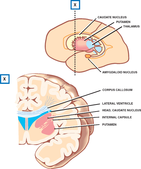
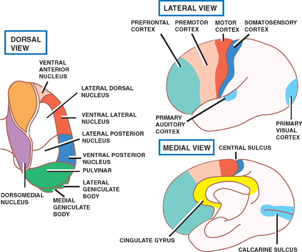
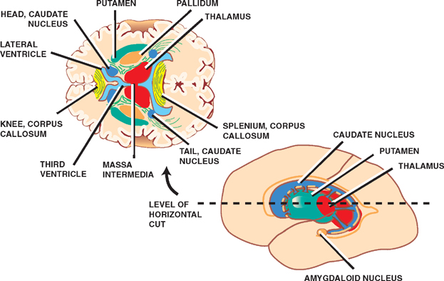
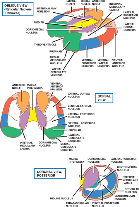
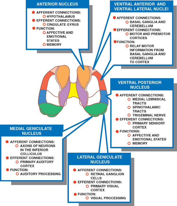
9 Thalamus The cerebrum comprises a central core, or diencephalon, surrounded by the cerebral hemispheres, or telen-cephalon. The diencephalon contains the third ventricle and adjacent structures, including (1) the thalamus, (2) the subthalamus, (3) the epithalamus, and (4) the hypothalamus. The boundaries of the diencephalon extend from the posterior commissure dorsally to the interventricular foramina ventrally. Dorsally it is bordered by the roof of the third ventricle; ventrally it is bordered, ventral to dorsal, by the optic chiasm, the infundibulum, and the mammil-lary bodies. The internal capsule comprises the lateral boundaries. See Figs. 9.1, 9.2, 9.3, and 9.4. The thalamus is a large, ovoid mass of gray matter situated on either side of the third ventricle. The medial surfaces of the thalami often meet within the third ventricle to form the interthalamic connection (or massa intermedia). The thalamus is the gateway to the cerebral cortex: all major sensory pathways, except olfactory, relay in the thalamus before ascending to the cortex. The thalamus also has important connections with the extrapyramidal motor system, the consciousness system, the visual system, and the limbic system. Therefore, lesions in the thalamus result in sensory and motor disturbances, as well as disturbances in alertness, vision, and behavior. The reticular nucleus excepted, each thalamic nucleus sends axons to the cerebral cortex. These terminate in either a discrete or a diffuse distribution. Fibers that are distributed discretely are precisely paralleled by reciprocal corticothalamic projections. In addition to the two-way cortical connections, other (subcortical) structures project afferents to the thalamus, and the thalamus projects efferents to the striatum (caudate and putamen) and hypothalamus. The reticular nucleus receives collaterals from both afferent and efferent thalamic axons and projects in turn to the other nuclei of the thalamus. No other thalamic nucleus projects to other thalamic nuclei, although each individual nucleus contains interneurons. In what follows, we first describe the structural organization of the thalamus and its nuclei, then describe a second classification based on function. After a brief description of thalamic vascular supply, we summarize the clinical manifestations that result from thalamic dysfunction. Fig. 9.3 Thalamic nuclei and their respective projections to the cortex. Fig. 9.4 Thalamus. See Fig. 9.5. Rostrally, the thalamus is covered by a thin white matter layer termed the stratum zonale. Laterally, it is covered by another white matter layer termed the external medullary lamina. The thalamus is divided into three main parts by a Y-shaped white matter layer termed the internal medullary lamina. The internal medullary lamina divides the thalamus into anterior, medial, and lateral aspects, the anterior portion lying between the limbs of the Y, the medial and lateral parts lying on either side of its stem. The lateral part of the thalamus is divided into two tiers: a dorsal tier that contains (1) the lateral dorsal nucleus, (2) the lateral posterior nucleus, and (3) the pulvi-nar; and a ventral tier that contains (1) the ventral anterior nucleus, (2) the ventral lateral nucleus, (3) the ventral posterior nucleus, and (4) the medial and lateral geniculate nuclei. Other thalamic nuclei not included in the three-part scheme are (1) the reticular nucleus, which is located on the lateral surface of the thalamus between the external medullary lamina and the internal capsule; (2) the intralaminar nuclei, which are located within the internal medullary lamina; and (3) the midline nuclei, which are located on the medial surface of the thalamus in the in-terthalamic connection. Fig. 9.5 Morphological classification of thalamic nuclei. See Fig. 9.6. There are three groups of functionally distinct tha-lamic nuclei: specific relay nuclei, nonspecific thalamic nuclei, and association nuclei. Each specific relay nucleus receives discrete input from a single sensory modality or a particular motor function and projects to a well-defined area of the primary sensory and motor cortex. Reciprocal corticothalamic connections faithfully copy each “specific” thalamocortical projection. The specific nuclei include the anterior nucleus and the ventral tier of the lateral nuclear group (ventral anterior, ventral lateral, ventral posterior, medial geniculate, and lateral geniculate nuclei).
Thalamus
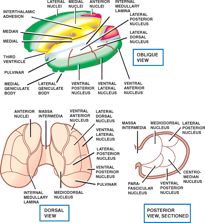
Morphological Classification
Functional Classification
Specific Relay Nuclei
Related posts:

Stay updated, free articles. Join our Telegram channel

Full access? Get Clinical Tree


