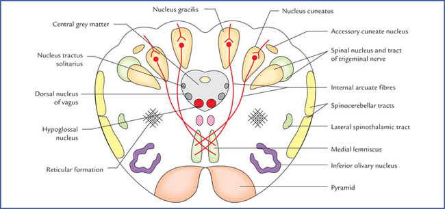
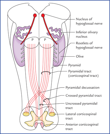
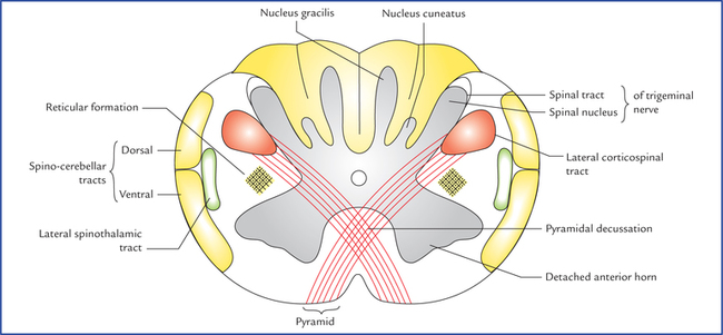
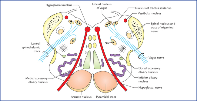
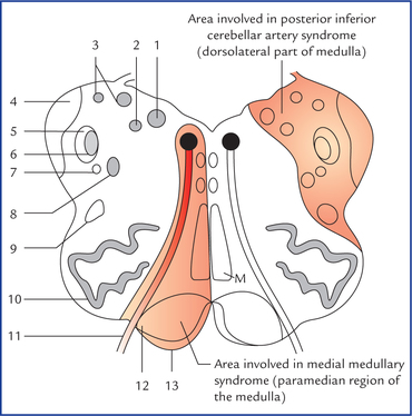
8 The brainstem is the stalk-like part of the brain which connects the spinal cord with the forebrain. From below upwards it consists of three parts: medulla oblongata, pons, and midbrain. The midbrain is continuous above with the cerebral hemispheres and the medulla oblongata is continuous below with spinal cord. Brainstem is located in the posterior cranial fossa. Its ventral surface lies on the clivus. Posteriorly, the pons and medulla are separated from the cerebellum by the cavity of the fourth ventricle. The brainstem serves the following four major functions: • It provides passage to various ascending and descending tracts that connect the spinal cord to the different parts of the forebrain. • It contains important autonomic reflex centres (vital centres) associated with the control of respiration heart rate and blood pressure. • It contains reticular activating system which controls consciousness. • It contains important nuclei of the last ten cranial nerves (i.e. IIIrd to XIIth). Medulla provides attachment to last four cranial nerves. An overview of structural components of medulla and their functions is provided in Table 8.1. Table 8.1 Structural components and functions of the medulla oblongata The medulla is divided into right and left symmetrical halves by anterior median fissure and posterior median sulcus (Fig. 8.1). Fig. 8.1 External features on the anterior (ventral) aspect of the brainstem. Note the attachment of last cranial nerves. Each half of the medulla is marked by two sulci— anterolateral and posterolateral, which are direct upward continuations of the corresponding sulci of the cord. The ventral aspect of medulla presents following features: • Pyramids. These are two elongated elevations, one on either side of anterior median fissure and are produced by the corticospinal (pyramidal) fibres. Most of these fibres about (75%) cross to the opposite side (pyramidal decussation) in the lower part of medulla and then descend as lateral corticospinal tract in the lateral white column of the spinal cord. About 20% of uncrossed fibres run downwards as anterior corticospinal tract in the anterior white column of the spinal cord; the remaining 5% run downwards along with uncrossed fibres in the lateral white column of the spinal cord (Fig. 8.2). Fig. 8.2 Showing decussation of pyramidal (corticospinal) tracts in the lower part of medulla oblongata and location of lateral and anterior corticospinal tracts in the spinal cord. (P = pyramid.) • Olives. These are oval elevations, posterolateral to the pyramids and are produced by an underlying mass of grey matter called inferior olivary nucleus. • Rootlets of the hypoglossal nerve. These emerge from the anterolateral sulcus between the pyramid and the olive. • Inferior cerebellar peduncles. These are thick bundles of fibres lying posterolateral to the olive, and attach the medulla with the cerebellum. • Rootlets of the IXth, Xth, and XIth (cranial part) cranial nerves. These emerge through the posterolateral sulcus separating the olive from the inferior cerebellar peduncle. The dorsal aspect the medulla is well demarcated into lower closed and upper open parts. Fig. 8.3 External features on the posterior (dorsal) aspect of the brainstem. Note the features in the floor of the fourth ventricle. The open part of the medulla forms the lower part of the floor of fourth ventricle, which presents number of features like, median sulcus, hypoglossal and vagal triangles, vestibular areas, area postrema, stria medullaris, etc. (Fig. 8.3). For details see floor of fourth ventricle on page 122. The section at this level passes through the inferior half of the medulla, and closely resembles to that of spinal cord. However, following important features are observed at this level (Fig. 8.4): Fig. 8.4 Transverse section through the lower closed part of the medulla oblongata at the level of pyramidal decussation. • The nucleus gracilis and nucleus cuneatus appear as the narrow strip like projections from the posterior aspect of the central grey matter. • The apex of posterior horn gets swollen up to form the nucleus of spinal tract of trigeminal nerve. It is an upward continuation of the substantia gelatinosa in the posterior grey column of the spinal cord. • The spinal tract of trigeminal nerve is a bundle of fibres which caps over the nucleus of spinal tract of trigeminal nerve. • Decussation of pyramidal tracts forms the most important feature of medulla at this level. About 75% fibres of pyramidal tract run backwards and laterally across the midline to reach the lateral white column of the opposite side of the spinal cord, where they run downwards as the lateral corticospinal tract. In doing so the anterior horns are detached from the central grey matter. • Each detached anterior horn divides to form the spinal nucleus of accessory nerve and supraspinal nucleus of first cervical nerve. The nucleus of accessory nerve extends downwards up to fifth cervical spinal segment. The supraspinal nucleus gives off the efferent fibres of the first cervical nerve, and is continuous above with the nucleus of the hypoglossal nerve. • Appearance of diffuse zone containing a network of fibres and scattered nerve cells within it the lateral white column adjacent to nucleus of spinal tract of trigeminal nerve is called reticular formation. This section passes through the middle of medulla and displays following features (Fig. 8.5): • The nucleus gracilis and nucleus cuneatus become more pronounced and are separated from the central grey matter. The fibres of fasciculus gracilis and fasciculus cuneatus occupy the broad posterior white column and terminate in these nuclei. • The internal arcuate fibres arising from the cells of gracile and cuneate nuclei (second order sensory neurons conducting sensations of discriminative touch, position and vibration) course forwards and medially around the central grey matter and decussate with corresponding fibres of opposite side in the median plane (sensory decussation) and then turn upwards to ascend as the medial lemniscus on the opposite side close to the median plane. In this decussation the gracile fibres are medial to that of cuneate fibres. • The internal arcuate fibres cut off the spinal nucleus and tract of trigeminal nerve from the central grey matter. • Immediately dorsolateral to the cuneate nucleus lies the accessory cuneate nucleus which receives the more lateral fibres (derived from the cervical segments of the cord) of the fasciculus cuneatus and gives rise to posterior external arcuate fibres conveying proprioceptive impulses to the cerebellum of the same side through inferior cerebellar peduncle. • The separated spinal nucleus and tract of trigeminal nerve lies ventrolateral to the cuneate nucleus. • The lower part of inferior olivary nucleus is seen. • The pyramids lie on either side of the anterior median fissure. • The central grey matter contains: (a) hypoglossal nucleus, (b) dorsal nucleus of vagus, and (c) nucleus of tractus solitarius. The hypoglossal nucleus occupies the ventro-medial position close to the midline in the central grey matter. The dorsal nucleus of vagus lies dorsolateral to the hypoglossal nucleus and nucleus of tractus soli-tarius lies just dorsolateral to the dorsal nucleus of vagus. • Medial longitudinal bundle lies posterior to the medial lemniscus. It is small compact tract of nerve fibres which interconnect the IIIrd, IVth, VIth, VIIIth and spinal nucleus of XIth cranial nerve nuclei. • Spinocerebellar and lateral spinothalamic tracts lie in the anterolateral area of lateral white column. • Lateral and anterior spinothalamic tracts are very close to each other and collectively form spinal lemniscus. Transverse section passes across the floor of the fourth ventricle and through the middle of olives and presents following features (Fig. 8.6): Fig. 8.6 Transverse section of medulla at the level of olives: 1. medial longitudinal fasciculus, 2. tectospinal tract, 3. medial lemniscus. (NA = nucleus ambiguus). • The central grey matter is spread over the floor of the fourth ventricle and contains the nuclei of several cranial nerves. From medial to lateral these are: hypoglossal nucleus, nucleus intercalatus, dorsal nucleus of vagus and vestibular nuclei (inferior and medial). • The nucleus of tractus solitarius lies ventral to vestibular nuclei. • The nucleus ambiguus lies deep within the reticular formation and gives origin to the motor fibres of IXth, Xth and XIth cranial nerves. • On either side of midline (paramedian region), from dorsal to ventral lie: medial longitudinal fasciculus, tectospinal tract, medial lemniscus, and pyramidal (corticospinal) tract. • The arcuate nuclei, thought to be inferiorly displaced pontine nuclei are situated on the anteromedial aspect of the pyramids. They receive fibres from the cerebral cortex and send efferent fibres to the cerebellum of the opposite side through the anterior external arcuate fibres. • Laterally, from dorsal to ventral lie two prominent structures: (a) inferior cerebellar peduncle, and (b) inferior olivary nucleus. 1. The inferior cerebellar peduncle occupies posterolateral part. 2. The inferior olivary nucleus is the largest mass of grey matter, and forms the most prominent feature in the section through upper part of medulla. It presents a crumbled bag like appearance. Close to the main nucleus lies medial and dorsal accessory olivary nuclei. The medulla is supplied by the following arteries: Vascular Disorders of Medulla Oblongata The common vascular lesions involving the medulla are the thrombosis of posterior inferior cerebellar and vertebral arteries leading to lateral and medial medullary syndromes respectively. Thrombosis of posterior inferior cerebellar artery therefore, affects a wedge-shaped area on the dorsolateral aspect of medulla (Fig. 8.7) and the inferior surface of the cerebellum and produces following signs and symptoms. Fig. 8.7 The transverse section of the upper part of the medulla. The red areas indicate the sites of lesions: 1. dorsal nucleus of vagus, 2. nucleus of tractus solitarius, 3. vestibular nuclei, 4. inferior cerebellar peduncle, 5. spinal tract of trigeminal nerve, 6. spinal nucleus of trigeminal nerve, 7. descending sympathetic tract, 8. nucleus ambiguus, 9. lateral spinothalamic tract, 10. inferior olivary nucleus, 11. hypoglossal nerve, 12. pyramidal tract, 13. arcuate nucleus. (M = medial lemniscus.) – Contralateral loss of pain and temperature sensation, in the trunk and limbs, due to involvement of spinothalamic tract. – Ipsilateral loss of pain and temperature sensation over the face, due to involvement of the spinal nucleus and tract of trigeminal nerve. – Ipsilateral paralysis of muscles of palate, pharynx and larynx due to involvement of nucleus ambiguous. – Ipsilateral ataxia, due to involvement of inferior cerebellar peduncle and cerebellum. – Giddiness, due to involvement of vestibular nuclei. – Horner’s syndrome, due to involvement of descending sympathetic pathway in the reticular formation of medulla.
Brainstem
Medulla Oblongata
Components
Functions
Grey matter
• Nucleus gracilis and nucleus cuneatus
Relay conscious proprioceptive sensations to the thalamus
• Olivary nuclei
Relay information associated with voluntary muscle movement to the cerebellum
• Vital centres
– Cardiac centre
Regulates heart rate and force of contraction
– Vasomotor centre
Regulates distribution of blood flow in vessels
– Respiratory centre
Regulates respiratory movements
• Nuclei of last four cranial nerves (IX, X, XI and XII)
See Chapter 9
• Other nuclei
Relay ascending sensory information from spinal cord to the higher centres
White matter
• Ascending and descending tracts
Connect the spinal cord with the other parts of the brain (to and fro)
External Features
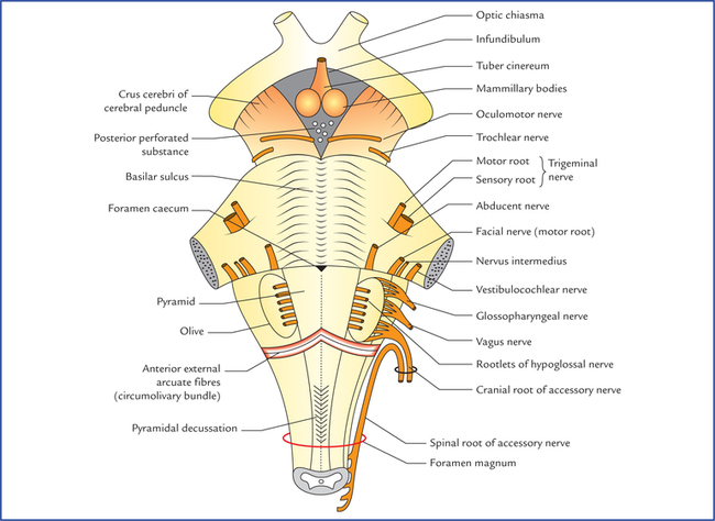
Features on the anterior (ventral) aspect of medulla (Fig. 8.1)
Features on the Posterior (Dorsal) Aspect of Medulla (Fig. 8.3)
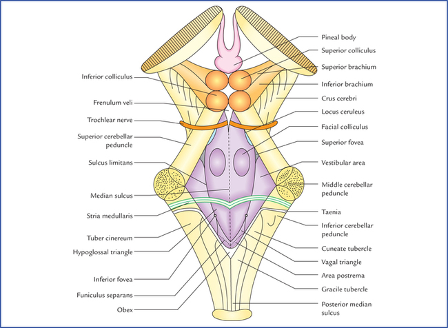
Features of the open part
Internal Structure
Transverse section of medulla at the level of pyramidal decussation (the great motor decussation)
Transverse section of medulla at the level of sensory decussation
Transverse section of medulla at the level of olives
Blood Supply of the Medulla
![]()
Stay updated, free articles. Join our Telegram channel

Full access? Get Clinical Tree