Cauda Equina Syndrome
Bryson Borg, MD
DIFFERENTIAL DIAGNOSIS
Common
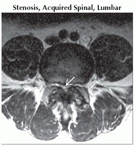
Stenosis, Acquired Spinal, Lumbar
Intervertebral Disc Herniation, Lumbar
Spondylolisthesis
Trauma
Burst Fracture, Lumbar
Sacral Fracture (Zone 3)
Traumatic Spondylolisthesis
Penetrating Injury
Post-Operative Spinal Complications
Abscess, Epidural, Paravertebral
Neoplasm
Metastases
Ependymoma, Myxopapillary, Spinal Cord
Meningioma
Arachnoid Cyst
Hematoma, Epidural-Subdural
Less Common
Multiple Sclerosis, Spinal Cord
Rare but Important
Sarcoidosis
Type IV AVF
Tethered Spinal Cord
Ankylosing Spondylitis
CIDP
Guillain-Barré Syndrome (Atypical Presentation)
ESSENTIAL INFORMATION
Key Differential Diagnosis Issues
Cauda equina syndrome (CES): Low back pain, sciatica, leg weakness, saddle hypoesthesia/anesthesia, urinary incontinence or retention, and incontinence of bowel
Substantial clinical overlap between the syndromes of the cauda equina and the conus medullaris
Most common cause is herniation of the intervertebral disc
MR or CT myelography are useful modalities to evaluate causes of CES
Acute onset CES generally considered a neurosurgical emergency, with best results if decompressed within 24-48 hours
Helpful Clues for Common Diagnoses
Intervertebral Disc Herniation, Lumbar
CES occurs in approximately 1-2% of cases of herniated lumbar disc
Most patients will have a long-standing history of back problems with or without unilateral sciatica
Post-Operative Spinal Complications
Misplaced pedicle screw
Displaced surgical device (fusion cage, graft material, artificial disc)
Epidural hematoma or abscess
Incomplete decompression
Retained sponge
Image Gallery
Nipple Structure
 Axial T2WI MR shows severe lumbar canal narrowing at L4-5
 secondary to disc bulge, ligamentous hypertrophy, and marked facet osteophyte. secondary to disc bulge, ligamentous hypertrophy, and marked facet osteophyte.Stay updated, free articles. Join our Telegram channel
Full access? Get Clinical Tree
 Get Clinical Tree app for offline access

|