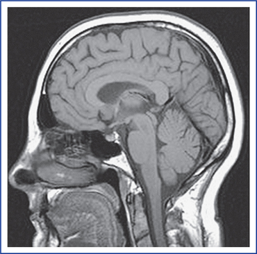
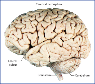
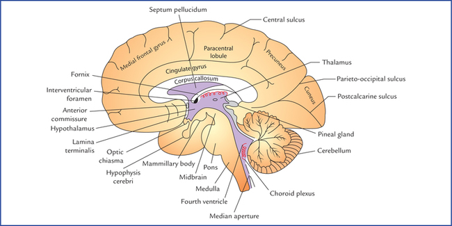
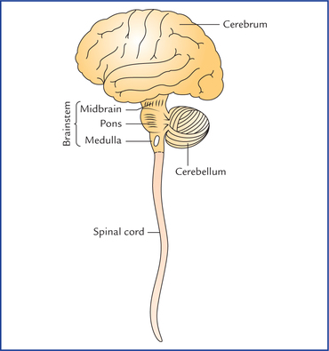
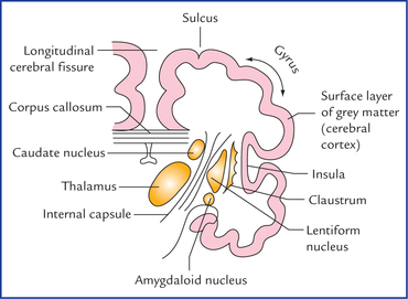
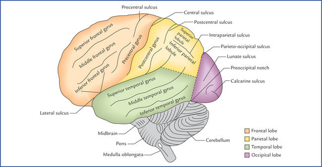
6 The central nervous system (CNS) consists of the brain and spinal cord, which are located in the cranial cavity and vertebral canal respectively (Fig. 6.1). The brain is that part of the CNS which lies within the cranial cavity. The functions of the brain are as follows: • It receives information from, and controls the activities of trunk and limbs mainly through its connections with the spinal cord. • It receives the information from, and controls the activities of head and neck structures through cranial nerves. • It assimilates experiences, a requisite to higher mental processes such as memory, learning and intelligence. • It is also responsible for one’s personality, thoughts and aspirations. The adult brain constitutes about one-fiftieth of body weight and weighs about 1400 g in males and 1200 g in females. It consists of six major parts: (a) the cerebrum, (b) the diencephalon, (c) the midbrain, (d) the pons, (e) the medulla oblongata, and (f) the cerebellum (Fig. 6.2). Fig. 6.2 The diagram showing parts of central nervous system. The diencephalon is not seen. The superior, middle and inferior cerebellar peduncles, which connect the cerebellum with the mid-brain, pons and medulla oblongata are shown schematically. The midbrain, pons and medulla oblongata collectively form the brainstem. The Figure 6.3 shows the superolateral aspect of the brain, as seen in dissection. Each cerebral hemisphere consists of a surface layer of grey matter, called cerebral cortex and a central core of white matter. In the basal part of the latter are located large masses of grey matter, known as basal nuclei/ganglia (Fig. 6.4). Fig. 6.4 A coronal section of the cerebral hemisphere showing surface layer of grey matter (cerebral cortex), central core of white matter and basal nuclei. The surface of cerebral hemisphere is convoluted, i.e. it has a series of elevations, the gyri, separated by shallow depressions, the sulci or deep grooves called fissures (Fig. 6.3). The superolateral surface of each cerebral hemisphere is divided into four lobes, which are named after the overlying skull bones (Fig. 6.5): Fig. 6.5 Superolateral surface of the brain. Note the demarcation of lobes on the superolateral surface of the cerebral hemisphere. • Frontal lobe is anterior to the central sulcus and above the lateral sulcus. • Parietal lobe is posterior to the central sulcus and above the lateral sulcus. • Occipital lobe is behind a line extending from parieto-occipital sulcus to the pre-occipital notch. • Temporal lobe is below the lateral sulcus and in front of parieto-occipital notch. The occipital lobe is responsible for reception and integration of visual input. The medial surface of the cerebral hemisphere is visualized in the sagittal section of brain and presents a number of features as shown in Figure 6.6. The basal ganglia include the lentiform nucleus, caudate nucleus, claustrum, and amygdaloid body (Fig. 6.4). During development of connections between the cerebral cortex and the brainstem, the bundles of fibres, converging as the internal capsule, partly divide the corpus striatum into a medial caudate nucleus and a lateral lentiform nucleus. Between the internal capsule and the cerebral cortex, the nerve fibres diverge as the corona radiata. The diencephalon is the part of brain between the cerebrum and the brainstem. Its main components are: (a) two thalami, (b) hypothalamus, (c) metathalamus, (d) epithalamus, and (e) subthalamus.
Central Nervous System: an Overview
Brain
Cerebrum
Basal ganglia/nuclei
Diencephalon
![]()
Stay updated, free articles. Join our Telegram channel

Full access? Get Clinical Tree


Central nervous system: an overview

