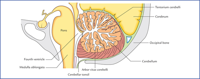
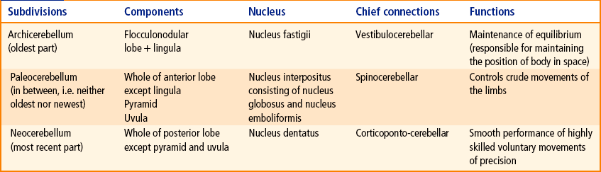
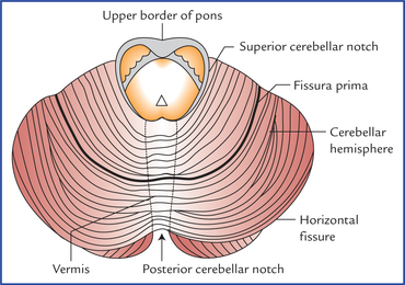
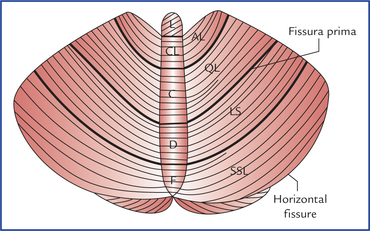
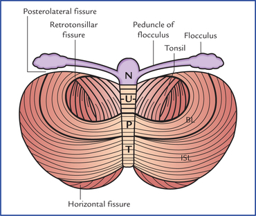
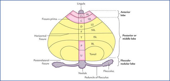
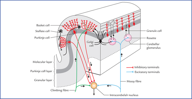
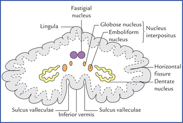
10 The cerebellum (L. cerebellum = little brain) is the largest part of the hindbrain and second largest part of the brain as a whole. It weighs about 150 g. It is located in the posterior cranial fossa underneath the tentorium cerebelli and behind the pons and medulla oblongata. It is separated from the pons and medulla by a cavity of the fourth ventricle (Fig. 10.1). Its surface bears numerous fissures separating narrow folia which are mostly transverse. The three primary functions of the cerebellum are: In addition to above, it also adjusts coordination of skilful volitional movements by perfect timing among the contracting groups of agonist and antagonist muscles. This is achieved through the use of somatic sensory information (proprioceptive sensations from muscles and joints) in modulating the motor output from the cerebrum and brainstem (see comparator function of cerebellum on page 119). Sherrington regarded the cerebellum as the head ganglion of the proprioceptive system. The cerebellar disease manifests the following triad of motor dysfunctions: • Dysequilibrium, i.e. loss of balance characterized by gate and trunkal ataxia. • Hypotonia, i.e. loss of the resistance normally offered by muscles on palpation. The external features of the cerebellum comprise three parts, two surfaces, two notches, and three well-marked fissures (Figs 10.2–10.4). Fig. 10.2 Superior view of the cerebellum. Note the presence of fissures and folia on the surface of the cerebellum. Fig. 10.3 Schematic diagram to show the various subdivisions of the cerebellum as seen on the superior surface. (L = lingula, CL = central lobule, C = culmen, D = declive, F = folium, AL = ala, QL = quadrate lobule, LS = lobulus simplex, SSL = superior semilunar lobule.) Fig. 10.4 Schematic diagram to show the various subdivisions of the cerebellum as seen on the inferior surface. (N = nodule, U = uvula, P = pyramid, T = tuber, BI = biventral lobule, ISL = inferior semilunar lobule.) The superior surface of the cerebellum is convex. The two cerebellar hemispheres are continuous with each other on this surface. The inferior surface presents a deep median notch called vallecula which separates the two cerebellar hemispheres. The floor of the vallecula is formed by inferior vermis and is limited on each side by sulcus valleculae. • The horizontal fissure is most conspicuous and runs along the lateral and posterior margins of the cerebellum. It marks the junction between the superior and inferior surfaces of the cerebellum. • The posterolateral fissure lies on the inferior surface of the cerebellum and separates the flocculonodular lobe from the rest of the cerebellum (corpus cerebelli). • The V-shaped fissura prima on the superior surface cuts the superior vermis at the junction of its anterior two-third and posterior one-third. It divides the corpus cerebelli into anterior and posterior (middle) lobes. N.B. There are several other fissures which subdivide the vermis and cerebellar hemispheres into lobules and given fanciful names (Table 10.1). Table 10.1 Various subdivisions (lobules) of vermis and cerebellar hemisphere Anatomically the cerebellum is divided into three lobes (Fig. 10.5), viz. anterior, posterior and flocculonodular. Fig. 10.5 Morphological and functional subdivisions of the cerebellum. The organ is being opened out (schematically) to show both superior and inferior surfaces together. The parts seen above the horizontal fissure form the superior surface and those below the fissure inferior surface of the cerebellum. (CL = culmen, C = central lobule, D = declive, F = folium, T = tuber, P = pyramid, U = uvula, AL = ala, QL = quadrate lobule, LS = lobulus simplex, SSL = superior semilunar lobule, ISL = inferior semilunar lobule, BL = biventral lobule.) • The anterior lobe lies on the superior surface anterior to the fissura prima. • The posterior/middle lobe lies between the fissure prima on the superior surface and posterolateral fissure on the inferior surface. • The flocculonodular lobe is smallest of all and lies on the inferior surface in front of the posterolateral fissure. The subdivisions (lobules) of the vermis and cerebellar hemispheres which constitute these lobes are listed in Table 10.1. The cerebellar cortex is folded in such a way that the surface of cerebellum presents a series of parallel transverse fissures and intervening narrow leaf-like bands called folia. Each folium consists of a slender branched lamina of central core of white matter covered by a thin layer of grey matter. The central core of white matter being arranged in the form of the branching pattern of a tree, is called arbor vitae cerebelli (arbor vitae = tree of life). The structure of the cerebellar cortex is uniform throughout (homotypical). Fig. 10.6 Structure of cerebellar cortex, showing intrinsic neurons and their processes. A single folium is shown cut longitudinally (right) and transversely (left). Note that cerebellar glomerulus is a synaptic complex involving four types of neurites: the mossy fibre terminal, the dendrites of granule and Golgi cells, and the Golgi cell axon terminal. The nerve cells are of two types: (a) the basket cells, and (b) the stellate cells. Purkinje cells layer consists of a single row of large flask-shaped cells, the Purkinje cells. A dendrite arises from the neck of the flask, passes upwards into the molecular layer, where it undergo profuse branching to form an elaborate dendritic tree. The dendrites of Purkinje cells synapse with: (a) the collaterals from the basket cells, (b) the axons of the granule cells (parallel fibres), and (c) the climbing fibres. The axons of the Purkinje cells arise from their deeper poles and pass through the granular layer into the white matter, where they relay into the intracerebellar nuclei (except those from the flocculonodular lobe which pass directly to the vestibular nuclei). The Golgi cells are large and prominent but scanty. Their den-drites ramify in the molecular layer. The intracerebellar nuclei (also called central nuclei) are masses of grey matter embedded in the white matter of the cerebellum. On each side of the midline they are four in number. From lateral to medial side these are: (a) dentate nucleus, (b) emboliform nucleus, (c) globose nucleus, and (d) fastigial nucleus (Fig. 10.7). Fig. 10.7 Horizontal section of the cerebellum showing intracerebellar nuclei (also called central nuclei of the cerebellum).
Cerebellum and Fourth Ventricle
Cerebellum
External Features
Surfaces
Fissures
Lobes
Subdivisions of vermis
Subdivisions of cerebellar hemisphere
Anterior lobe
Lingula
Central lobule
Culmen
No lateral projection
Alae
Quadrangular lobule
Posterior lobe
Declive
Folium
Tuber
Pyramid
Uvula
Lobulus simplex
Superior semilunar lobule
Inferior semilunar lobule
Biventral lobule
Tonsil
Flocculonodular lobe
Nodule
Flocculus
Subdivisions of Cerebellum
Anatomical subdivisions
Internal Structure
Grey matter
Structure of cerebellar cortex (Fig. 10.6)
Molecular (plexiform) layer
Purkinje cell layer
Granular layer
Intracerebellar nuclei
![]()
Stay updated, free articles. Join our Telegram channel

Full access? Get Clinical Tree


Cerebellum and fourth ventricle