Chapter 19 Chest and Abdominal Wall
Thoracic Ventral Spinal Rami
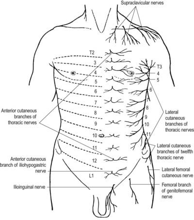
There are 12 pairs of thoracic ventral rami. The upper 11 lie between the ribs (intercostal nerves), and the twelfth lies below the last rib (subcostal nerve) (Figs 19.1, 19.2). Each is connected with the adjoining ganglion of the sympathetic trunk by grey and white rami communicantes; the grey ramus joins the nerve proximal to the point at which the white ramus leaves it. Intercostal nerves are distributed primarily to the thoracic and abdominal walls. The first two nerves supply fibres to the upper limb in addition to their thoracic branches, the next four supply only the thoracic wall and the lower five supply both thoracic and abdominal walls. The subcostal nerve is distributed to the abdominal wall and the gluteal skin. Communicating branches link the intercostal nerves posteriorly in the intercostal spaces, and the lower five nerves communicate freely in the abdominal wall.
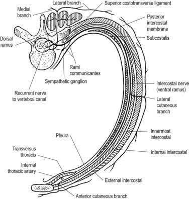
Fig. 19.1 Course of a typical intercostal nerve. The muscular and collateral branches are not shown.
First to Sixth Thoracic Ventral Rami
The second to sixth thoracic ventral rami pass forward in their intercostal spaces below the intercostal vessels. At the back of the chest they lie between the pleura and external intercostal membranes, but in most of their course they run between the internal intercostals and the subcostals and innermost intercostals (see Fig. 19.2). Near the sternum, they cross anterior to the internal thoracic vessels and transversus thoracis; pierce the internal intercostals, external intercostal membranes and pectoralis major; and end as the anterior cutaneous nerves of the thorax, which supply the skin on the front of the thorax. The second anterior cutaneous nerve may be connected to the medial supraclavicular nerves of the cervical plexus; twigs from the sixth intercostal nerve supply abdominal skin in the upper part of the infrasternal angle.
Branches
The lateral cutaneous branch of the second intercostal nerve is the intercostobrachial nerve (see Fig. 18.9). It crosses the axilla to gain the medial side of the arm and joins a branch of the medial cutaneous nerve of the arm. It then pierces the deep fascia of the arm and supplies the skin of the upper half of the posterior and medial parts of the arm, communicating with the posterior cutaneous branch of the radial nerve. Its size is in inverse proportion to the size of the medial cutaneous nerve. A second intercostobrachial nerve often branches off from the anterior part of the third lateral cutaneous nerve and sends filaments to the axilla and the medial side of the arm.

Stay updated, free articles. Join our Telegram channel

Full access? Get Clinical Tree


