Neuromuscular Junction

Chapter 22 Neuromuscular Junction


Skeletal Muscle


The most intensively studied effector endings are those that innervate muscle, particularly skeletal muscle. All neuromuscular (myoneural) junctions are axon terminals of somatic motor neurones. They are specialized for the release of neurotransmitter onto the sarcolemma of skeletal muscle fibres, causing a change in their electrical state that leads to contraction. Each axon branches near its terminal and subsequently innervates from several to hundreds of muscle fibres, depending on the precision of motor control required. The detailed structure of a motor terminal varies with the type of muscle innervated. Two major endings are recognized: those typical of extrafusal muscle fibres, and endings on the intrafusal fibres of neuromuscular spindles. In the former, each axon terminal usually ends midway along a muscle fibre in a discoidal motor end-plate (Figs 22.122.3). This type usually initiates action potentials, which are rapidly conducted to all parts of the muscle fibre. In the latter, the axon has numerous subsidiary branches that form a cluster of small expansions extending along the muscle fibre. In the absence of propagated muscle excitation, these excite the fibre at several points. Both types are associated with a specialized receptive region of the muscle fibre, the sole plate, where a number of muscle cell nuclei are grouped within the granular sarcoplasm.
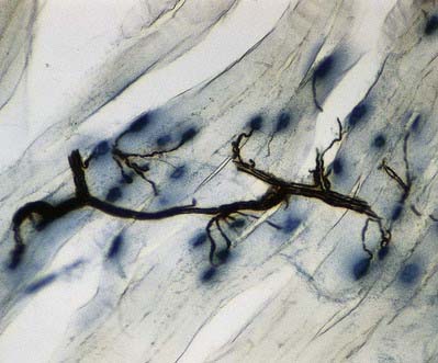
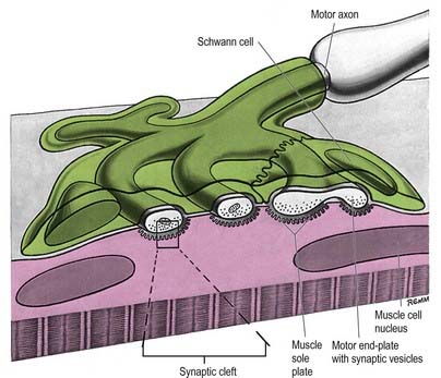


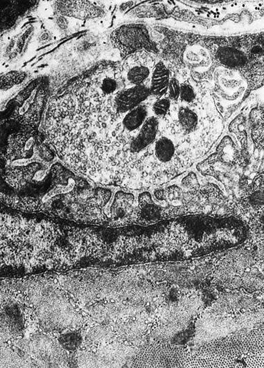
The sole plate contains numerous mitochondria, endoplasmic reticulum and Golgi complexes (see Figs 22.2, 22.3). The neuronal terminal branches are plugged into shallow grooves in the surface of the sole plate (primary clefts), from which numerous pleats extend for a short distance into the underlying sarcoplasm (secondary clefts). The axon terminal contains mitochondria and many clear 60-nm spherical vesicles, similar to those in presynaptic boutons, clustered over the zone of membrane apposition. The motor terminal is ensheathed by Schwann cells whose cytoplasmic projections extend into the synaptic cleft. The plasma membranes of the nerve terminal and the muscle cell are separated by a 30- to 50-nm gap, with a basal lamina interposed. The basal lamina follows the surface folding of the sole plate membrane into the secondary clefts. It contains specialized components, including specific isoforms of type IV collagen and laminin and agrin, a heparan sulphate proteoglycan. Endings of fast and slow twitch muscle fibres differ in detail: the sarcolemmal grooves are deeper, and the presynaptic vesicles more numerous, in the fast fibres.


Junctions with skeletal muscle are cholinergic, and the release of acetylcholine (ACh) changes the ionic permeability of the muscle fibre. Clustering of ACh receptors at the neuromuscular junction depends in part on the presence of agrin, synthesized by the motor neurone. Agrin affects muscle cytoskeletal attachments to the ACh receptor cytoplasmic domain and prevents their lateral diffusion out of the junction. When the depolarization of the sarcolemma reaches a particular threshold, it initiates an all-or-none action potential in the sarcolemma, which is then propagated rapidly over the whole cell surface and also deep within the fibre via the invaginations (T-tubules) of the sarcolemma, causing contraction. The amount of ACh released by the arrival of a single nerve impulse is sufficient to trigger an action potential. However, because ACh is very rapidly hydrolysed by the enzyme acetylcholinesterase (AChE), present at the sarcolemmal surface of the sole plate, a single nerve impulse gives rise to only one muscle action potential—that is, there is a one-to-one relationship between neural and muscle action potentials. Thus, the contraction of a muscle fibre is controlled by the firing frequency of its motor neurone. Neuromuscular junctions are partially blocked by high concentrations of lactic acid, as in some types of muscle fatigue.


Conduction of the Nervous Impulse


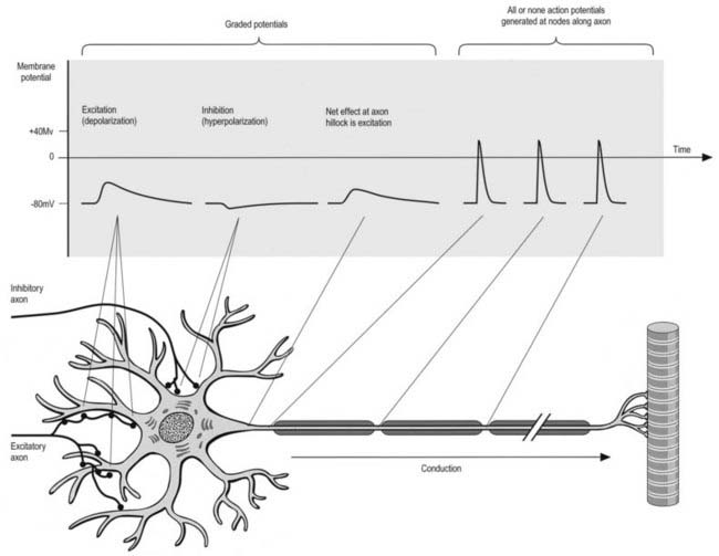
All cells generate a steady electrochemical potential across their plasma membranes (a membrane potential) because of the different ionic concentrations inside and outside the cell (Fig. 22.4). Neurones use minute fluctuations in this potential to receive, conduct and transmit information across their surfaces.


The membrane potential of a neurone, known as the resting potential, is similar to that of non-excitable cells. In most neurones it is approximately 80 mV, being negative inside. The entry into neurones of sodium or, in some sites, calcium ions causes depolarization of the cell, whereas an increased chloride influx or an increased potassium efflux results in hyperpolarization. Plasma membrane permeability to these ions is altered by the opening or closing of ion-specific transmembrane channels, triggered by chemical or electrical stimuli. Chemically triggered ionic fluxes occur at synapses and may be either direct, whereby the chemical agent (neurotransmitter) binds to the channel itself to cause it to open, or indirect, whereby the neurotransmitter is bound by a transmembrane receptor molecule that is not itself a channel but that activates a complex second messenger system within the cell to open separate transmembrane channels. Electrically induced changes in membrane potential depend on the presence of voltage-sensitive ion channels that, when the transmembrane potential reaches a critical level, open to allow the influx or efflux of specific ions. In all cases, the channels remain open only transiently, and the numbers that open and close determine the total flux of ions across the membrane.


The types and concentrations of transmembrane channels and related proteins, and therefore the electrical activity of the membranes, vary in different parts of the cell. Dendrites and neuronal somata depend mainly on neurotransmitter action and show graded potentials, whereas axons have voltage-gated channels that give rise to action potentials.


In graded potentials, a flow of current from or into adjacent areas of the cell occurs when a synapse is activated, and this contributes to the total degree of polarization of the membrane covering the cell body. However, the influence of an individual synapse on neighbouring regions decreases with distance, so that, for instance, synapses on the distal tips of dendrites may, on their own, have relatively little effect. The electrical state of a neurone therefore depends on many factors, including the number and position of thousands of excitatory and inhibitory synapses, their degree of activation, the branching pattern of the dendritic tree and the geometry of the cell body. The target of these integrated factors is a small part of the neurone surface, the axon hillock, where voltage-sensitive channels are concentrated (unlike the dendrites or somata). The axon hillock is the site where action potentials are generated before being conducted along the axon.


CASE 1 Botulism


A 22-year-old woman presents in the early morning with double vision, slurred speech and swallowing difficulties, preceded the night before by dry mouth, blurred vision, abdominal cramping and constipation. During the next several hours, she develops limb weakness and finally shortness of breath requiring intubation. She has otherwise been well. However, she participated in a covered-dish dinner at a family reunion on the day the symptoms began. Several other family members who attended also complained of abdominal cramps and diarrhea or constipation, and later the same day her mother experienced double vision and slurred speech.


On examination, she exhibits eyelid ptosis and complete bilateral ophthalmoplegia. Her pupils fail to react to light. She has marked facial and tongue weakness. Strength is reduced throughout, with absent reflexes.


Discussion: Botulinum toxin ingestion in adults initially causes gastrointestinal symptoms, followed by weakness, especially of ocular and bulbar muscles. Because the toxin’s site of action is presynaptic, decreased or lack of ACh release occurs at all peripheral cholinergic nerve endings, both nicotinic (neuromuscular junction) and muscarinic (autonomic) receptors. The toxin is taken up into the nerve terminal by endocytosis, then cleaves proteins required for the docking and fusing of vesicles to the presynaptic member, thereby blocking exocytosis and release of ACh. The binding of the toxin is permanent and results in destruction of the axon terminal. Regrowth of the nerve terminal must occur for function to return. Those affected early after ingestion usually have a more devastating course than those affected later. Clustering of cases frequently occurs.

Stay updated, free articles. Join our Telegram channel

Aug 14, 2016 | Posted by in NEUROLOGY | Comments Off on Neuromuscular Junction

Full access? Get Clinical Tree

Get Clinical Tree app for offline access