4 Clinical Picture
Leon Sazbon and Giuliano Dolce
General Considerations
Patients in the vegetative state (VS) are placed in specialized recovery units after an initial period of days or weeks in reanimation or intensive care. Before the individual medical diagnostic and therapeutic procedures are discussed, the general approach to management needs to be elucidated.
Prevailing medical practices in general apply to specific circumstances that warrant specific and predictable procedures. However, in patients in VS, some aspects of care necessarily diverge from these principles, and the protocol is in several ways independent of the patient’s clinical condition. For example, laboratory tests are usually not done unless specifically indicated, while roentgen films are made of all eight major joints during the first 6 months, to allow early detection of periarticular osseous formations. Because it is impossible to formulate a prognosis or predict either the clinical progression or the duration of the VS, the goals of management are twofold: to keep the patient alive and anticipate all possible complications; and to facilitate conscious activity – an indispensable condition for functional recovery. The fundamental rules guiding each step in patient treatment are precise, punctilious adherence to schedule (hours and days of selected interventions), order, and silence. These provide the patient with the requisite therapeutic milieu, minimizing the risk of potentially life–compromising, emergency conditions that can be induced by the grave clinical condition itself.
The clinical picture of the patient in VS consists of two interrelated parts: the internal symptomatology, which is unique in each individual case; and the universal neurological signs and symptoms and their changes over time. Progression of the first can affect the progression of the second. We will describe both of these aspects separately.
Medical Aspects and Complications
The complications and sequelae of VS form a wide spectrum of clinical pictures, affecting practically all biological systems. Medical, neurological, and surgical problems can complicate the rehabilitation process and even pose a threat to life. Some are a direct consequence of the alteration in brain function and its autonomic and hormonal influences [1], and others are nursing–related or iatrogenic. This section describes the most common complications of VS in relation to the organ or system affected.
Cardiovascular Effects
Cardiovascular complications are more frequently observed in the acute phase following head trauma, when the patient is still comatose, although they may carry over into the rehabilitation phase of VS. The rate of patients affected by cardiovascular complications varies from 7% [2] to 32% [3].
Hyperdynamic cardiovascular reactivity to head trauma is manifested by three main clinical symptoms: neurogenic hypertension, changes in cardiac rate, and arrhythmia.
• Moderate degrees of systemic hypertension (160 mmHg systolic or more) in the absence of high intracranial pressure is a frequent finding [4]. Kaliski et al. [3] reported hypertension in 11 % of 180 patients with severe head injuries and a mean coma duration of 55 days.
• Changes in cardiac rate are frequently observed. Tachycardia occurs more often than bradycardia, which is a poor prognostic sign [5].
• Among the arrhythmias, ventricular extrasystoles are seldom recorded. Sazbon and Grosswasser [2] and Kaliski et al. [3] also described electrocardiographic findings of nonspecific S –T and T wave changes.
According to Kirland and Wilson [6], the hypothalamus and nucleus of the solitary tract are the brain areas implicated in this type of hyperdynamic cardiovascular response. In addition, patients in VS with associated chest trauma may suffer myocardial damage, with cardiac contusion and subendocardial hemorrhage.
Respiratory Complications
Patients in VS may show normal or periodic respiratory rhythms. Among the latter, it should be emphasized that the presence of central hyperventilation, a well–recognized pattern of periodic breathing, indicates a bad outcome [7]. Another type of periodic respiratory rhythm is ataxic breathing, an absolutely irregular respiratory pattern, which is also associated with very poor prognosis; and adult respiratory distress syndrome, which was observed by Sazbon and Groswasser in 7% of patients [2].
A certain degree of hypoxia, with Po2 between 60 and 80 mmHg in arterial blood, combined with a certain degree of hypercarbia, is the rule. The coexistence of these two conditions is characteristic of inadequate ventilation due to lung obstruction, lung collapse, bronchospasm, pulmonary edema, mechanical failure, or drug overdose [8,9]. Eisenberg and Levin [10] claimed that in acute comatose patients, the presence of the triad of high intracranial pressure, hypoxia, and hypotension following brain trauma may predict either VS or death. In patients who recover from VS, tidal volume is normal, but both the inspiratory and expiratory phases of vital capacity are markedly reduced, as is forced vital capacity [11].
Tracheobronchial infection or colonization is an almost unavoidable complication in patients who have undergone intubation or tracheostomy. Tracheal granulation also occurs very frequently, which can lead to stenosis. Tracheomalacia, tracheoesophageal fistula, and innominate artery erosion are severe but very rare complications. Parenchymal infections were reported in 37% of 134 patients in one series of patients in VS for at least 1 month, but pulmonary abscess is rare. Atelectasis, fat embolism related to concurrent limb fracture in the earliest weeks, and pulmonary embolism following deep phlebothrombosis, are not uncommon. Macrobronchial aspiration is often fatal, and recurrent microaspiration leads to pneumonia.
Rib fractures and unilateral concomitant traumatic paralysis of the phrenic nerve are cofactors of hypoventilation and recurrent pulmonary problems.
One iatrogenic respiratory complication is pneumothorax, caused by a failed attempt at subclavian vein cannulation.
Associated finger clubbing, unilateral or bilateral, has been frequently observed [12].
Urinary Complications
Lack of sphincter control is an important clinical feature of trauma-induced VS. Clinicians must perform tonus bladder studies to avoid overlooking a neurogenic bladder, which can lead to upper urinary tract impairment. Krimchansky et al. [13] studied bladder function in 17 patients in VS for 1 month and found that all of the patients had neurogenic bladder of the hypertonic type. There were no cases of detrusor – sphincter dyssynergia or unstable bladder. Some authors have tentatively claimed that it is the pons that controls detrusor and external sphincter function [13,14], whereas others localize this function to the precentral lobe of the cortex [15].
The urinary tract is often the site of hospital–acquired infection in patients with VS, with reported incidences of 39% [2] and 37% [3]. Most cases are associated with the use of an indwelling catheter or intermittent catheterization; the longer the duration of catheterization, the higher the risk [1]. In patients with an indwelling catheter, asymptomatic bacteriuria is common; fever, when it occurs, may be due to the urinary infection or another source, and the reason is often hard to determine. Treatment of asymptomatic bacteriuria is strongly discouraged.
The use of absorbent pants or diapers may be associated with maceration of the skin. Penile inflammation, erosions, or ulcerations are also usually related to external sources.
Surprisingly, urolithiasis is very rare (2%), despite the often long–term bedridden status of these patients [16].
Gastrointestinal Complications
Gastrointestinal complications develop in 50% of patients in VS [3]. There is often bleeding from the digestive tract, both in the acute phase and even years after onset. In most cases, the hemorrhage is microscopic, and esophagitis, gastritis, or ulcers may be found on gastroesophageal endoscopy [17]. The mechanism remains unclear [16], since after several weeks and months stress can be excluded as an underlying factor. Gastric pH is usually normal. In patients with gastric acidity, neutralization with antacids or H2–blockers can cause bacterial overgrowth, which may be a risk factor for pneumonia. Reflux is another major complication, given that all patients in VS are fed by nasogastric tube or percutaneous endoscopic gastrostomy. Fluoroscopic studies show that in the supine position, rapid introduction of the food bolus causes regurgitation and sometimes, in turn, bronchial aspiration. Reflux may be avoided by slowing the food delivery and placing the patient in a reclining (semisitting) position. Gastric reflux is not related to the dietary components, food quantity, or presence of a feeding tube [18].
Patients in VS often have impaired liver function [3]. Biopsy series have reported contradictory results. Aiges et al. [19] noted no liver cell damage or inflammatory reactions, but this finding was not supported by Kaliski et al. [3]. The elevation in liver enzymes may be a consequence of either viral hepatitis following blood transfusion (often done in the acute phase after trauma) or, in the absence of a positive hepatitis serologic screen, administration of anticonvulsant or other drugs. Although vegetative patients are incontinent, constipation due to inactivity is common. At the same time, diarrhea may be caused by hyperosmolar foods or overgrowth of bacteria, particularly Clostridium difficile; its severity varies widely.
Motor Problems
The pathology of head injury is heterogeneous, involving diffuse axonal injury, multifocal cortical contusions, and secondary hypoxic damage. This may lead to disorders of movement and to disturbances of motor control and tonus [20].
Spasticity
Upper motor neuron (pyramidal) lesions may manifest clinically as spasticity – that is, hyperreflexia of the stretch reflex, resulting in a velocity–dependent increase in tendon reflexes [21]. The reported rate of spasticity in a Japanese series of VS patients was 32% [22]. Its biological basis is complex and not well understood. Current opinion holds that spasticity derives from an interruption of the corticospinal, vestibulospinal, and reticulospinal tracts and their modulatory influences on the spinal alpha motor neurons [23]. Symptoms may be positive, such as exaggerated nociceptive reflexes and withdrawal, autonomic hyperreflexia, dystonia, and contractures, or negative, such as paresis and fatigability [24]. In bedridden patients, the long-term inactivity can lead to shortening and retraction of the tendons and joint deformities and contractures. Other possible sequelae are bedsores, respiratory infections, osteoporosis, sepsis, deep vein thrombosis, pulmonary embolism, and periarticular new bone formation. Patients in VS may adopt diverse postures, but generally, progressive limb flexion is seen, either in abduction or adduction; the fingers are flexed, with the thumb hidden under the other fingers or emerging between the second and third finger. In rare cases, there may be extension of one or more fingers, usually the second or the fifth; clawing is noted even less often. The head shows a tendency to lean backward owing to hypertonus of the paravertebral muscles, occasionally achieving a position of opisthotonos.
Rigidity
Rigidity is a cardinal sign of extrapyramidal syndrome. Lesions of the basal ganglia in VS have been described by several authors (see Chapter 3). In the Japanese series presented by Higashi et al. [22], rigidity was noted in 62 % of patients. Jennett and Teasdale [25], in an autopsy study, noted ischemic lesions in 91% of head–injury patients, 89% of whom had lesions of the basal ganglia. Bricolo [26] and Graham et al. [27] reported that half of the patients in VS had hemorrhage or necrosis of the basal ganglia as a consequence of direct trauma, in addition to ischemic lesions of the putamen and pallidum. In rare cases, there are involuntary movements (hemiballism, chorea or athetosis) and/or disorganized movements. Generally, muscle hypotony is very frequent until the end of the first month of VS, when it begins to decrease gradually while tonus increases, ultimately to the characteristic flexor hypertonus, sometimes accompanied by isolated isometric muscular contractions.
Plegia or Paresis
Plegia or paresis is almost the rule in VS. Feldman [28], in an unpublished series of 117 patients in VS of traumatic origin for at least one month, found a 70 % rate of tetraplegia and a 30 % rate of hemiplegia. Higashi et al. [22] found that 55 % of their patients in VS were tetraplegic, 18 % were hemiplegic, and the remainder were triplegic or monoplegic.
Motor Reactivity
Abnormal involuntary movements or normal uncontrolled movements are the result of extensive cerebral lesions causing disturbances of sensory input or of processing or programming by the motor areas. Examples are sucking, chewing, or swallowing, pelvic crural flexion movements, scratching, and slow, search–like movements.
Sazbon and Groswasser [4], in a study of 99 patients in VS, reported a 13 % rate of flaccid motor unresponsiveness –67% decerebrate reactivity and 15% decorticate reactivity. Normal motor reactivity was seen in only 4% of the patients [4]. A mixture of decerebrate and decorticate reactivity is not uncommon in individual patients (e. g., a decerebrate response on one side of the body or in one limb and a decorticate one on the other side, or in the rest of the limbs).
Periarticular New Bone Formation
Periarticular new bone formation (PNBF) is due to abnormal metaplasia of the connective tissue near the joints, in which undifferentiated mesenchymal cells undergo transformation under the influence of unknown factors and form histologically true bone. The reported frequency in VS varies from 11 % to 76% in various studies [29–34], probably owing to differences in selection procedures. It is usually noted at the time of a transversal cut, or reported retrospectively. In a prospective radiological of patients in VS for 2–32 months, Sazbon et al. [30] found that PNBF is an expression of a progressive disease, starting about 1–2 months after trauma and peaking at up to 5 months. The process is self–limited, terminating at 14 months or before. PNBF is mainly localized to the large joints, mostly in the shoulder, followed by the hip, and then the elbow and knee. Hand and spinal involvement is rare [35,36]. The clinical picture is divided into three types: pseudoinflammatory, pseudotumoral, and ankylosing. The direct cause remains obscure. Suggested possible etiologic factors, all based on scanty evidence, include long–term immobility, spasticity, paralysis, trophic changes in the affected limb, local trauma to the affected joint, or traumatic effect of physiotherapy [29–31,37]. Sazbon et al. [30] found no correlation between the presence of PNBF and mechanical ventilation in the early stage of coma or recurrent genitourinary infections, and no correlation between its progression and the patient’s age or sex, or with the etiology, duration, or outcome of VS. The appearance of PNBF may be identified by a specific L-dopa test and a nonspecific thyrotropin-releasing hormone test, with an increase in growth hormone concentrations in both [38]. PNBF undoubtedly poses a major problem during comprehensive rehabilitation programs.
PNBF is not specific to VS. It accompanies a variety of neural and extraneural insults, of which traumatic lesion of the brain or spinal cord is the most common. It may also be related to poliomyelitis, tetanus, carbon monoxide poisoning, burns, and stroke [38].
Seizures
Epilepsy
Epilepsy is a temporary brain dysfunction consisting of recurrent, unprovoked hyperchronous discharges of cortical neurons [39,40]. Post-traumatic epilepsy is characterized by recurrent seizure episodes in patients with brain injuries that cannot be attributed to any other cause [41]. The reported incidence of post-traumatic epilepsy varies from 1.9% to 53% [3,37,42–55]. Specifically, in VS, Higashi et al. [22] and Feldman [28] both reported an incidence of almost 50%, whereas Sazbon and Grosswasser [4] reported only 32 %. The occurrence of epilepsy varies with age (with a greater likelihood in younger adults) [46,55], genetic predisposition, type of injury (penetrating or closed), duration of unconsciousness [39,52,53], formation of intracranial hematomas of any type, presence of intracranial infection or depressed skull fractures, and tearing of the dura mater [27,54]. The epileptic attacks may occur early after the traumatic event (from a few seconds to up to 1 month after), or later (delayed epilepsy). In patients with closed depressed skull fractures, Jennett et al. [55] found no differences in the incidence of late epilepsy between patients who underwent elevation of the fragment bone and those receiving conservative treatment. The attacks may be focal or secondary and generalized; the latter type is more persistent over time. Caveness et al. [56] reported that 25 % of their severely brain–injured patients had focal attacks and 50% had both – either separately, or secondary and generalized following focal.
Myoclonus
Myoclonus refers to involuntary rough and very brief muscular contractions, with or without displacement of body parts. The presence of myoclonus usually indicates a metabolic cause or a severe anoxic episode complicating the primary traumatic injury to the brain [57]. Johnston and Sheshia [58] and Snyder et al. [45] reported that myoclonic seizures have an even worse influence on vital and functional outcome than generalized epilepsy. Myoclonus may appear, like generalized seizures, as bilaterally synchronous myoclonic twitches [59] or, more frequently, as status epilepticus. Two variants are seen in VS patients: opsoclonus–myoclonus, defined as rapid chaotic movement of the eyes accompanied by myoclonus of the limbs and cerebellar signs [60]; and palatal myoclonus, defined as rhythmic contractions of the soft palate due to a lesion of the olivary nuclei in the medulla oblongata [61]. It is often discovered by the rhythmic “jumps” of the nasogastric tube, which rests on the palate. This disorder is sometimes accompanied by jerks of other muscled structures deriving from the same branchial arch – for example, the pharynx, larynx, tongue, face, orbicularis oris, and neck [62]. At times, it is triggered by sensory stimuli – even mild ones such as a gentle touch [63].
Ventricular Enlargement
Post-traumatic hydrocephalus or ventricular enlargement is considered a frequent complication or sequela of severe brain injury. The reported incidence varies widely, from 0.7 % to 62 % [64–70]. In VS specifically, studies note incidences of 37% [3] and 51 % [2]. The dilatation may be widespread, involving the whole ventricular system, or may be limited mainly to the lateral ventricles (central atrophy) or cortical sulci (cortical atrophy). According to Kaliski et al. [3], the reported range of ventricular dilatation depends on the method used to determine ventricular size.
Post-traumatic ventricular dilatation may be due to wasting of the white matter (ex vacuo hydrocephalus), adhesions of the meninges and circulatory impairment of the cerebrospinal fluid (obstructive hydrocephalus), or malabsorption of the cerebrospinal fluid (normal–pressure hydrocephalus) [25]. Radiologically, empty images of the porencephalic type may be seen when they communicate with cerebrospinal fluid (CSF) passages [26]. It is particularly important to determine whether the ventricular enlargement is due to cerebral atrophy (ex vacuo) or to a communicating hydrocephalus, because there is no treatment for the former, whereas the latter requires ventricular shunting (see Chapter 6). In patients in VS, communicating hydrocephalus cannot be recognized clinically, as the classic diagnostic triad of dementia, incontinence, and gait disturbances is absent. The decision on whether to perform shunt procedures is based on the clinical course (i. e., arrest or regression in the clinical course), and on progressive enlargement of the ventricles, with ballooning of the third ventricle, without clear, dominant brain atrophy or massive loss of brain tissue.
Undernutrition
Severe brain trauma induces several metabolic changes leading to severe weight loss, often in the order of 50% of the previous weight. In VS, cachexia is frequent [26,71–73], with an incidence of 20% to 48% [2,22]. The weight loss is due to the elevated energy expenditures and increased catabolism characteristic of the acute phase of severe brain injury. This abnormality may continue for several months, into the stage of VS. In addition to the effects of neuroendocrine and autonomic alterations, the increased cardiac output, hyperventilation, fever, restlessness, posturing, seizures, and infections impose a metabolic burden [1]. Patients with abnormal posturing reactivity have the highest consumption of energy. When no nutritional support is provided, there is a large protein and caloric deficit and depletion of fat stores. This, in turn, leads to loss of immunocompetence, depression of defense mechanisms against infection [74], and hindrance of internal homeostasis, injury repair, and wound healing, and ultimately to a state of marasmus and cachexia. Calorie and protein supplementation, in the order of 40–60 cal/kg/day, drastically reduces mortality [75].
Water and Electrolyte Balance
Conditions of depletion or excess of water and electrolytes may appear separately, but a mixed syndrome is more common. Water constitutes approximately 60% of the total body weight, with two–thirds being intracellular and the rest consisting of plasma and interstitial fluids. Potassium is the predominant intracellular cation, and sodium the predominant extracellular cation; organic acids are the major intracellular anion, and chloride the major extracellular anion. In VS, there may be alterations in serum potassium, calcium and phosphorus concentrations, but more frequent and relevant are sodium disturbances – both hypernatremia and hyponatremia.
Hyponatremia and Hypernatremia
Hyponatremia has been related to permanent brain damage [76,77]. Among the syndromes involved are inappropriate antidiuretic hormone secretion (IADHS) syndrome and water intoxication [78]; the latter is usually iatrogenic. IADHS has been reported in 8.2% [79] and 12.3% [4] of braindamaged patients. Dilutional hyponatremia may be caused by drugs such as thiazides, mannitol, barbiturates, and carbamazepine [80,81]. Endocrinological dysfunctions such as Addison’s disease have also been implicated, but an acute etiologic diagnosis requires an evaluation of the extracellular fluid as well. In an unpublished work, Sazbon [82] suggested that for the clinical diagnosis of hyponatremia and hypo-osmolarity, the biceps should be pinched firmly between thumb and forefinger and quickly released; in affected patients, a thick, distinct band of muscle will be noted, which lasts for a few seconds. Signs of dehydration may be seen in cases of reduced extracellular fluid.
Hypernatremia is also often iatrogenic. It results from inadequate water replacement in the presence of an excess of water–to–sodium loss. The most representative syndrome is diabetes insipidus, due to lack of pituitary antidiuretic hormone secretion. Causes include exaggerated diuresis provoked by high protein tube feeding, gross sodium intake, or excessive water loss due to profuse sweating or diarrhea. There may also be water loss through the respiratory tract in patients who have undergone tracheostomy, and increased insensible water loss during periods of fever.
Hypokalemia and Hyperkalemia
Hypokalemia is more frequent than hyperkalemia. It can be due to gastrointestinal losses, large therapeutic use of laxatives, decreased dietary potassium intake, and loss of potassium in sweat. Other causes are corticosteroids or diuretics, or a hormonal imbalance. In vegetative patients, hypokalemia leads to muscular weakness, which may in turn lead to respiratory failure, paralytic ileus, and cardiac arrhythmia.
Hyperkalemia is associated with acidosis, renal failure, oliguric states, administration of potassium-sparing diuretics or exaggerated potassium supply. The most serious side effect is cardiac toxicity.
Other
Calcium, magnesium, and phosphorus disorders are seldom seen in VS. True hypocalcemia, when it occurs, is related to a reduction in protein–bound calcium due to hypoalbuminemia. It is asymptomatic, since membrane excitability is produced by the ionized fraction, which is unaltered. Hypomagnesemia is related to prolonged parenteral nutrition in combination with gastrointestinal losses or inadequate intake. Clinical manifestations are weakness, neuromuscular hyperexcitability, tremor, and changes in the electrocardiography (ECG) findings. Hypophosphatemia may be the result of the use of large amounts of aluminum hydroxide antacid, or may be part of the nutritional recovery syndrome.
Hematological Alterations
Diffuse intravascular coagulation (DIC) occurs as a consequence of a major intravascular activation of the coagulation cascade. In severe forms of brain injury, thromboplastin is released into the blood circulation from the injured zones [6,83–86], activating the extrinsic pathway of the clotting cascade. The actual incidence of DIC in VS is unknown, but it is likely very infrequent relative to its high frequency in the acute period of injury. Perpetuation of the syndrome has been reported in rare cases [87]. In spite of the decreased number of platelets, low level of fibrinogen, and abnormal values of plasma split products, clinical manifestations are extremely rare.
Anemia in VS is most often the “anemia of chronic or inflammatory diseases.” It may also be due to dietary deficiencies of iron, vitamin B12, or folic acid, or to drug sensitivity hemolysis in cases of glucose–6–phosphate dehydrogenase (G6PD). Although a mild degree of gastrointestinal bleeding is very frequent in VS, it is rarely the cause of anemia.
Hormonal Disorders
Hypothalamic – pituitary dysfunction may be manifested clinically as hypothyroidism, hypoadrenalism, hypopituitarism, diabetes insipidus, or IADHS. Hypopituitarism and diabetes insipidus are well recognized hormonal complications of the acute stage of head injury. In rare cases, they evolve into serious, life–threatening conditions; in others, minor degrees of hypothalamic – pituitary dysfunction may be present without clear, related symptomatology, especially in the presence of severe unconsciousness that may mask the clinical signs [88–100]. In VS, symptoms may begin a few hours after trauma or only days, months or years later. These include persistent low body temperature, low pulse and blood pressure, hypoglycemia, hyponatremia, and changes in diuresis volume. Slightly diminished levels of cortisol, testosterone, luteinizing hormone, follicle–stimulating hormone, triiodothyronine (T3), thyroxine, and thyroid-stimulating hormone have been reported [5]. Sack et al. [101] suggested that a lack of reciprocity between T3 and rT3 may follow defective deiodination in both the alpha and beta rings of thyroxine. Precocious puberty, with elevated levels of 17–ketosteroids, and hyperthyreosis are unusual [5].
Sazbon and Groswasser [4] reported a 12% incidence of clinical signs of posterior lobe dysfunction in a series of 130 vegetative patients, and Bakay and Wood [102] noted transient blood hypo-osmolarity secondary to IADHS in two–thirds of their comatose patients. Daniel and Treip [103] reported hemorrhaging in the posterior lobe in 45 % of fatal brain injuries, and Kornblum and Fischer [94] in 62%. All the suggested mechanisms of injury involve denervation of the posterior hypothalamus. Kornblum and Fischer [94] noted that injury to the stalk may be fatal, whereas Porter and Miller [104] emphasized the role of traumatic elongation of the stalk. The hypophysis sits in the sella turcica at the base of the cranium and is well protected by its bony sheath [89]. Some authors have reported that the culprit lesion is confined to the hypothalamus [92–96,100]. However, Crompton [88], in an autopsy study, reported that lesions of the hypothalamus occur together with lesions of the pituitary gland, especially in young males, and that they are often associated with a temporoparietal impact. They consist of microhemorrhages due either to venous engorgement subsequent to increased cranial pressure, or directly to elevated intracranial pressure. Ischemic necrosis can occur anywhere in the hypothalamus – pituitary axis.
The relative immunity of the anterior lobe [5,89,98,102] may be explained by its high location and lack of participation in the hypothalamopituitary portal system [105].
Immunologic Disorders
Humoral deficits, neutrophil dysfunction, a defective reticuloendothelial system, and depressed cellular immune function have all been documented in severe brain injury [106–108]. Hoyt et al. [109] reported a decreased proliferative response of T–cells to mitogen stimulation, with unaffected B–cells [109]. Wollaj et al. [106] noted impaired humoral immunity in three of 11 patients in VS. Hemolytic activity of the classical pathway was depressed in association with low levels of C1q, C1r and C4. One patient also had very decreased levels of immunoglobulin G2 (IgG2) and IgG4, which substantially influenced the phagocytic arm. Neutrophils showed impaired killing activity secondary to the humoral defect. Chemotaxis, random migration, and superoxide release were normal.
Infections
Patients in VS are particularly susceptible to nosocomial infections. Risk factors include the use of indwelling catheters, endotracheal tubes or cannulas, and nasogastric tubes, undernutrition, diminished immunologic defenses, immobility, atelectasis, lack of normal cough reflex, and bedsores. Rates range from 34% to 46% [3,4,22,110]. The most common nosocomial infections are pneumonia, urinary tract infection, sinusitis, and pseudomembranous colitis; the most common culprit organisms are Pseudomonas and Klebsiella. Meningitis, subdural empyema, and brain abscess occur rarely (1–2% of cases) following evacuation of delayed hematoma, shunt procedures, or cerebrospinal fluid fistula [2,3,111]. They are usually caused by Staphylococcus aureus, S. epidermidis, Escherichia coli, Klebsiella, and Acinetobacter. Pyogenic arthritis is very rare [112].
Deep Vein Thrombosis
Deep vein thrombosis (DVT) is a frequent complication in severely brain–injured patients [113], with a reported incidence of 20–54% [113–115]. The most frequent location is the calf, followed by the intrapelvic and humeral or subclavian veins. There are no statistics on DVT in VS, although this patient group is characterized by several important risk factors: long duration of bed confinement, long–term immobility, and motor deficits; some patients have undergone neurological, abdominal, or orthopedic operations, or have leg injuries and Gram–negative sepsis. In addition, free fatty acid mobilization, which is a typical response to head injury to increase energy needs [116], has a known tendency to cause DVT due to hypercoagulation [117]. DVT and its complication of pulmonary embolism is detectable with 125I fibrinogen scanning, venous Doppler ultrasonography, and impedance plethysmography.
Autonomic Disturbances
Autonomic disorders are another well–recognized acute complication of traumatic brain injury. Symptoms may persist for months, even into VS.
Fever
Fever (hyperpyrexia) is extremely common in VS. In addition to such known causes as infections, other etiologies need to be considered as well – for example, PNBF, DVT, and drug reaction. The latter is found in 3–5% of cases, and is often related to hypersensitivity reactions [118]. Hypopyrexia due to hypothalamic damage is rare [4,57]. The diagnosis of fever of central origin is made by exclusion when a comprehensive work–up fails to yield a detectable cause. The reported incidence ranges between 4% and 7% [118,119]. Central fever is due to hypothalamic dysfunction. Its mediation by prostaglandin is controversial, although treatment with prostaglandin blockers such as indomethacin has been found beneficial. The fever is resistant to antipyretic drugs, but responds quickly to external cooling.
Sweating
Generalized sweating was noted in 25% of cases in one Israeli series [4]. In the absence of fever or other detectable causes, sweating indicates a failure of the hypothalamic thermoregulatory system. However, the underlying cause of bouts of localized sweating in the face and neck, often associated with flushing [4,71,120,121], is unclear.
Dysautonomia
Some authors consider dysautonomia, or vegetative storm, to be a distinct clinical entity affecting patients with severe traumatic brain injury [122,123]. It is characterized by a simultaneous and paroxysmal increase in at least five of the following seven autonomic parameters [123]:
• Heart rate (tachycardia > 120 beats/min)
• Respiratory rate (tachypnea > 30 breaths/min)
• Muscle tone (increased)
• Posture (decerebrate or decorticate)
• Blood pressure (systolic blood pressure > 160 mmHg)
• Sweating (profuse)
• Temperature (increased or decreased)
Episodes may occur with varying frequencies and times, or at specific hours of the day. They usually disappear within 4 months of the onset of unconsciousness. A diencephalic epileptic origin has been suggested.
| Characteristics | VS with dysautonomia | VS without dysautonomia |
| Patients (n) | 26 | 44 |
| Mean age (years) | 28.3 | 29.3 |
| Admission CCS | 6.7 | 7.7 |
| Days in ICU for VS patients | 168.5 | 85.6 |
| Syndrome duration (days) | 151 | – |
| Outcome | ||
| Moderate disability | 11.50% | 38.60% |
| Severe disability | 30.70% | 25.00% |
| VS > 1 y | 38.40% | 20.40% |
| Death | 19.20% | 15.90%* |
* CCS, Glasgow Coma Scale; ICU, intensive-care unit.
In a study of 70 patients in VS, Baguley et al. [123] found dysautonomic syndrome in about 30% of cases. According to Milano and Leto [122], the final functional outcome in affected patients is worse than that in patients without this syndrome. Their findings are summarized in Table 4.1.
Dermatological Changes and Bedsores
Acne vulgaris, seborrheic dermatitis, folliculitis, flat keloid changes, contact dermatitis, and allergic rash have all been reported in VS [2,3]. Patients are also prone to decubitus ulcers due to immobility, maceration of the skin due to contact with urine and feces, local sweating, and undernutrition. These may be exacerbated by deficient nursing care. Common sites of bedsores are the sacrum and trochanter region, heels, and occipital skin. Secondary complications of bedsores include localized osteoporosis, osteomyelitis, or osteoarthritis. Bedsores, in turn, may be a source of sepsis.
Complications of Medications
Drugs used in the treatment of patients in VS include anticonvulsants, benzodiazepines, beta blockers, antibiotics, L–dopa, and bromocriptine, among others, Skin reactions, angioedema, urticaria, fever, hematological changes, blood dyscrasias, and hepatic and renal toxicity may occur immediately after drug administration or later, with succeeding doses [124].
Other
Sharpening of the distal part of the fingers, finger clubbing, hypocratic fingers, hyperpigmentation of the dorsal aspect of the interphalangeal and metacarpophalangeal joints, ulnar deviation of the hands, longitudinal striation of the nails, and loss of hair are frequent findings, but of secondary value.
Neurological Aspects
Giuliano Dolce and Leon Sazbon
The neurological picture of the vegetative state has been described by several authors, mostly during the 1960s. In his book Das traumatische apallische Syndrom, Gerstenbrand described a number of detailed characterizing neurological signs [125], and included descriptions published by earlier authors, mostly in German or French [126]. These reports share a common fault: they contain an almost endless list of symptoms (mostly providing completely redundant information), but define poorly their sequence in time and their category of importance –possibly because most patients did not survive long enough. This refined exercise in neurological semiotics was curtailed by the appearance of emergency care and reanimation units/departments, and then by the development of brain imaging techniques for diagnosis. A list of neurological signs is given on p. 33–35. In our experience, however, we have always favored a standard neurological examination alongside morphological and functional examinations and brain imaging. As a result, we feel entitled to reduce expectations and above all to recognize the importance of numerous signs in the diagnostic and prognostic clinical evaluation (see p. 32–33).
Neurological Examination
The purpose of the neurological examination is to estimate the degree and level of functional organization of the patient’s nervous system, at least once every 2 weeks until signs of initial recuperation appear. The conscious interaction between the patient and the observer that is required in conventional neurological examinations is lacking in the vegetative state. The examination is therefore limited to observations of spontaneous activity, reflexes, and pathological phenomena involving primitive reflexes, which are quite numerous and various in this type of patient. The patient should be supine and undressed for this functional examination. In this section, each step of the neurological examination will be described in full detail in relation to the inspection and observation of complex activities, reflexes, and reactivity.
Posture
The alignment of the body and head and the positioning of the limbs are observed. Hyperextension to opisthotonos of the head and trunk are rare, whereas flexion of the head and upper body or lateral deviation of the head are observed more frequently. Each limb (or pair of limbs) can assume its own posture: the upper limbs are mostly flexed (decorticate posture) or hyperextended (decerebrate posture) [7,127]. When decerebration and decortication occur in association, it is possible to observe a limb or a whole side of the body in flexion while the other side is in extension. The lower limbs are mostly in hyperextension, but flexion of one or both limbs is not exceptional. Wrists and fingers are usually in flexion and pronation, while the feet generally assume plantar flexion and supination. None of these postural positions have particular prognostic value when considered individually, and they can revert completely within the first 3–4 months of the vegetative state. In contrast, tonic and postural disturbances may become permanent after this period, with associated deficits that make functional recuperation more difficult.
Spontaneous and Pathological Movements
It is necessary to observe and describe in detail all subtle, segmental movements, as well as the presence of pathological movements such as clonic and myoclonic tremors [128–130]. Spontaneous movements of flexion and extension of each and any of the four limbs or of single parts of a limb (such as the body rotation often observed in the first weeks or months) constitute a favorable sign for recovery of conscious activity. Conversely, the complete absence of spontaneous movement of the four limbs after the third or fourth month of vegetative state is a poor prognostic indication of a vegetative state progressing towards a recuperation of conscious activity (see p. 32–33).
Among the various forms of spontaneous pathological movement, decerebrate and decorticate crises are common in the early months, but revert completely or occur only rarely after the eighth to tenth weeks. Tremors, clonus, and myoclonic and dystonic crises are clinical expressions of specific neurological injury to the cortex and underlying subcortical structures of the brainstem and cerebellum and their pathways. They often appear early in the first weeks, with total or partial later remissions, and manifest differently in each individual depending on the location and type of damage incurred. These deficits have no particular value in the evaluation of the course of the vegetative state. They cannot stand in the way of the resumption of conscious activity, but may reduce, to a greater or lesser extent, the recovery of motor functions.
Passive Movements and Muscular Tone
Passive mobilization of limb segments and large and small joints allows the evaluation of the muscular tone and the identification of ossifications, periosteomas, and retractions of individual joints. These observations are particularly important in the vegetative state in order to help ascertain whether the absence of spontaneous movements results from paralysis or from mechanical impediments, such as the frequent blocked joints.
When limited to a single limb or limb segment, disturbances of the muscular tone such as flaccidity or spasticity may have different significance in relation to the general muscular tone as expressions of local function. They are therefore important for evaluating actual conditions, especially when marked by sudden or rapid changes. Marked hypertonia, with all limbs hyperextended, reflects functions at the mesencephalic level. Sudden progression to flaccidity of all limbs does not signify amelioration, but rather a regression to a bulbar level not exceeding the pons. In general, sudden changes occur in the early stages of vegetative state, or when the patient is still in coma, due to compression, wedging, or edema of the mesencephalic structures. Progression to diffuse flaccidity or to hypertonus is more common.
Disturbances of the muscular tone are quite frequent and constitute a serious obstacle to functional recovery as well as a considerable therapeutic task (see p. 76). All changes in muscular tone can revert, sometimes totally within the first 3 months, and are generally preceded by a recovery of conscious activities. After this period, a therapy–resistant widespread spasticity is generally observed, which may become a serious complication and a very unfavorable condition for the recovery of conscious mental activity, by limiting or impeding the resumption of movements that permit exploration of space.
Behavioral Responses
Following inspection, the examination of the patient begins with an attempt to establish communication, when and if possible. The patient is supine and with the eyes open. The gaze is empty and glassy, although wakeful; eye movements can be conjugated, but are not finalized to fixate or follow objects moving in the visual field. Patients do not answer to their name, nor can they respond to simple verbal commands or requests for imitation. This first part of the examination must be carried out from both sides of the patient.
Contact with the outer world, environment or people – even the closest relatives – is completely absent [4,131,132]. No significant “refusal” motor response, such as closure of the eyelids or opening of the jaws, nor any defense mechanisms are evident, with a resistance indicative of a lack of complex motility involving active cognition. A quantitative scale exploring different dimensions of the patient’s communication is useful to monitor the patient’s progress over time (see page 46). The use of the Glasgow Coma Scale in this condition may be inappropriate, as it matches the diagnostic and prognostic requirements of a different clinical entity – i. e., coma.
The “behavioral” examination of the patient in the vegetative state requires attention, dedication and experience in the observation and interpretation of every sign potentially conveying relevant information. Often, even apparently insignificant or nonspecific signs can provide useful information or indications of a certain degree of conscious activity. Opening of the mouth is not necessarily just a primitive, automatic action, but at times can be the only practicable way to express a need (”I am hungry, I am thirsty”) or to communicate feelings such as “I am surprised, I don’t agree, help me, I don’t feel well,” etc. It is therefore also necessary to consider the context in which certain actions occur before they are deemed to be automatic motor actions unrelated to cognitive function [133]. Each individual sign, once observed by the whole team and discussed on scheduled rounds, can then be assigned a certain value.
Ocular Motility
Ocular motility is examined with the patient’s head being kept in axis. Closed eyelids must be opened by applying intense pain stimuli to the skin of the clavicular fossa, first on one side and then the other. When the eyelids remain closed, the presence of local lesions or impairment of the oculomotor nerve must be checked for. In some cases, the patient forces his or her eyes shut, but this is not an act of volition. The same applies to the blink reflex to menace, which is sometimes absent at a first observation and may appear at a later time without signifying any integrated higher brain functions (such as those implied in the recognition of danger).
Spontaneous or provoked eye movements make it possible to note the presence of conjugated eye movements. Impairment of cranial nerves III and VI, possibly caused by peripheral lesions resulting from fractures and compressions at the base of the skull or orbits, must be recognized: monocular and binocular (horizontal, vertical, rotary, bobbing or retractive) nystagmus can be observed to occur either spontaneously or in response to passive head movements [7,134,135]. The presence of a bilateral nystagmus, in any more or less persistent form, is to be interpreted as the functional expression of massive anatomical damage at the mesencephalic level. It is hardly susceptible to regression, due to the likely involvement of the longitudinal medial bundle and reticular formation or of the centers regulating vigilance – which gives nystagmus a negative character.
It is not possible to evaluate a deficit of the fourth cranial nerve actively.
Pupils are observed for isocoria/anisocoria and isocyclicity, monolateral or bilateral miosis or mydriasis, and direct and consensual reflexes to light [136]. Accommodation cannot be tested in the absence of collaboration. The ciliospinal reflex, elicited by intense, painful stimulation of the neck or upper trunk skin, induces mydriasis. This reflex is almost always observed in the vegetative state (consequently excluding a deficit of the sympathetic cerebral chain in this condition), but can be absent in patients with concomitant direct trauma of the neck [137]. When observed in response to strong emotional stimulation, such as a visit from a special person, mydriasis may indicate that the afferent paths of Budge stemming indirectly from the hippocampus are unaffected.
The corneal reflex – i. e., closure of eyelids in response to a light touch on the cornea – makes it possible to evaluate the anatomical integrity of the afferent (trigeminal) pathway and motor response, in relation to the facial nerve and bulbopontine loop [43]. It is important to observe whether the corneal stimulus induces a corneo–chin reflex, a pathological response ascribed to the liberation of a cortical inhibition reflex of bulbopontine structures. A corneomandibular reflex (through contraction of the ipsilateral pterygoid muscle) can also be elicited in response to corneal stimulation and expresses cortical inhibition over a wider portion of the brainstem and bulbomesencephalic loop. The latter two reflexes are often observed in the vegetative state and constitute a characteristic indication of functional disconnection between cortical control and the brainstem (inhibitory function).
Oral Reflexes and Automatism
The “bulldog”, tactile-oral, oculo-oral, labiomental, and snout reflexes and the chewing, sucking or swallowing automatisms (either spontaneous or provoked by stimulation of the lips or oropharyngeal cavity) are almost pathognomic of both the post-traumatic and non – post-traumatic vegetative state and are, in any event, expressions of diffuse brain damage.
Reflexes and automatisms are expressions of the level of functional arrangement attained by the postlesional brain organization. The presence of some or all reflexes/automatisms takes on significance in the light of time of appearance, duration, and remission. From a pathophysiological point of view, the functional area or anatomical level able to elicit reflexes or automatic activity is important to evaluate. Only the oculo-oral reflex, although somewhat rare, expresses an enlargement of the functional area, including involvement of diencephalic structures, such as the lateral thalamic geniculate body, its connection with the mesencephalic roof, and the quadrigeminal bodies. The result is a functional integration of higher level in a sequence including the retina, thalamus, mesencephalic roof, motor nucleus of the fifth cranial nerve, and reticulospinal tract inducing also body flexion. This condition describes a notable enlargement of the reflexogenic area. Continuous mastication, like other motor automatisms, expresses an enlargement of the reflex area, involving the loss of a certain degree of selectivity, indicating a globalization and extension of the functional deficit. The cervical myotatic reflex, or head-retraction reflex, which is elicited by the percussion of the upper lip while the head is kept slightly flexed, also has an analogous meaning and must be recognized. The involved area is large and involves the bulb, pons, and cervical medulla. This reflex is rare, and when it occurs it is usually observed after 3–6 months of vegetative state. Another characteristic sign involving the musculature controlling mastication and closure of the mouth is the rabbitsnout phenomenon, which may be seen when the patient bites his or her lower lip – at times even biting off a piece of the lip [5]. This phenomenon always occurs after about 6 months and constitutes a late negative prognostic sign. After this period, the so–called half–moon pucker mouth can be seen, which is also a very negative prognostic sign.
Oculocephalic Reflex
The patient is supine, with the head in line with the trunk, and the head is quickly rotated laterally by the examiner (the reflex is elicited by movements of the head on the horizontal plane in patients in the vegetative state, due to the presence of a tracheal cannula limiting the head flexion). The eyes should follow the direction of the head. In the absence of the oculocephalic reflex observed in concomitance with brainstem lesions the eyes do not follow the direction of the head. The result is the so–called “doll’s eye sign” [138].
Trunk and Limbs
The patient’s control of the head and chest is indispensable to elaborate a treatment plan to be initiated in the early phase (see p. 97), and needs to be evaluated as soon as possible. The patient should be in a sitting position, with proper support (the erect posture is generally impossible). An additional and more elaborate evaluation of head and body control must be carried out with the patient prone or lying on either side. The patient should also be observed erect while supported by a special lifting or standing device (Fig. 4.1 and 7.4), and the presence of automatic step mechanisms should be checked for; rehabilitation procedures can take advantage of this function to facilitate motility of the lower limbs.
Motility of the limbs should be favored after the assessment of reflexes and only after having excluded impediments in the joints. For this purpose, automatic movements of portions of limbs from a painful stimulus applied to the skin can be evoked.
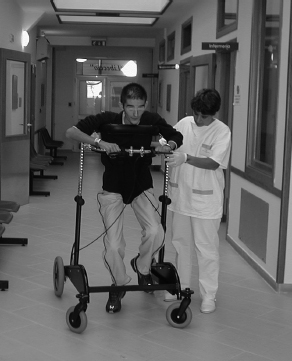
Fig. 4.1 The equipment used to position a patient in the vegetative state in an upright position, reducing the weight loading on the legs and feet and helping facilitate the motor scheme of walking
If present, these movements indicate that sensory functions are conserved to some degree.
Reflexes
Attenuated tendon reflexes are a functional correlate of compromised neuronal structures, without reflecting any functions peculiar to the vegetative state. Palm-chin and thumb-chin reflexes (physiological if observed until the third month of life) are always present in diffuse cerebropathies. At the beginning of vegetative state, the reflexogenic area can be enlarged to the extent that this reflex can be elicited by merely stroking the skin of the forearms and even the lateral parts of the trunk. This phenomenon expresses the functional liberation of structures of the brainstem and medulla from cortical control. This reflex can accompany head flexion and may be an indication of the level of functional organization of the cerebral structures.
Recovery of Conscious Activity
Recovery from the vegetative state occurs in different phases, notably: awakening with recovery of conscious activity, recuperation of functions through motor rehabilitation, readaptation based on the development of residual functions, and finally reintegration into family life and eventually into the working world.
These steps imply a neurological and psychological progression. During this entire period, the patient needs medical and social guidance in order to attain his or her objectives and must be assisted by a multidisciplinary team together with the family.
Patients usually progress from coma to the vegetative state while still in the emergency department. This progression is marked by two important steps – notably the patient’s opening of the eyes with a watchful look (at first in response to painful stimuli, then spontaneously) and the subsequent appearance of sleep–wake cycles in long–term electroencephalography (EEG) recordings. Transient or pseudorhythmic EEG waveforms peculiar to slow–wave sleep are initially observed, together with spikes at the vertex and K complexes. The sleep patterns become progressively more organized and finally also include periods of rapid eye movement (REM) sleep. For most of the time, sleep cycles are short in duration and alternate with periods of wakefulness. Only after several weeks can longer periods of nocturnal sleep be observed (see p. 67).
The progression from the vegetative state to an adequate recovery of conscious functions, inappropriately described as “awakening” or “reawakening,” is a pivotal moment in the patient’s clinical progress. For such a progression to come about, functional organization must occur at levels of complexity comparable to those required for the progression from coma to vegetative state. The opening of the patient’s eyes correlates functionally to a reestablished functional organization of the brain (or more precisely of the brains-tem structures), in which the nuclei of the cranial nerves regulating ocular globe and eyelid activity, as well as the part of the reticular formation in which the activating system originates, act together and simultaneously (arousal). It is therefore among these structures that a new functional organization allowing the resumption of conscious activity emerges. This is characterized by better control achieved by the brainstem structures partly substituting for the damaged cortex and by a higher degree of reciprocal interaction between these systems.
Clinically, this renewed organization of cerebral structures is expressed by a corresponding modification of neurological functions. The oral automatisms (such as chewing, sucking, swallowing) and pathological reflexes (such as the bulldog reflex or the oculo-oral and corneal-chin reflexes) disappear. Spontaneous mobility (if previously absent) reappears. A cardinal sign that anticipates the resumption of the most elementary conscious activities also appears–a conjugated “human” gaze following sources of visual stimulation (tracking) and oriented towards changes in the visual field induced by moving people or objects. This sign is an expression of functional integration at high levels of complexity, implying a recognition of form and significance of objects or people, and above all, indicating the complex motor activity required for spatial exploration. Thereafter, the ipsilateral and contralateral primitive motor systems resume function, together with the resumption of thalamocortical circuits through the pallidal system, in the absence of tectal reflexes. These neurological signs precede the resumption of conscious activity and are necessary prerequisites for this process to occur.
It is at this stage that the patient emerges from the vegetative state (awakening without awareness). The speed and quality of recuperation depend on the degree of residual functional plasticity. This factor is peculiar to each individual patient and linked to age, with the most extensive recuperation being observed in young patients 25–30 years old or younger. The recuperation of conscious functions presents very differently from case to case due to the involvement of many factors, the most relevant of which is undoubtedly the patient’s own personality before the accident that led to the vegetative state.
Qualitative aspects, as well as the time necessary to stabilize functions, are diverse and do not always depend on small changes, but rather on individual curves of progress, without any correlation with the location and extension of the brain lesion. In all cases, the ability to execute simple commands is evident first, with the patient using an indifferent action (such as closing the eyes, opening of the mouth, or squeezing the hand) that can vouch for a certain function or response. The second level attained is defined as “reactive consciousness” [139], a term covering the state that lasts until the appearance of consciousness of the outer world. Then comes a phase defined by increasing interchange and interaction with the surroundings, which leads to recuperation of a certain degree of consciousness of the self.
The first stage ends when the patient is regularly able to follow simple commands such as closing the fist or eyes or opening the mouth in a predictable manner. This can last for weeks or months and does not make it possible to predict any progression into the next phase.
Gaze movements, unfinalized motor actions, yawning, chewing, and differentiated mimicry (expressing suffering or well–being) can arise and may be used to establish a primitive relationship with the patient and suggest a possible progression into successive phases. However, an established relationship with the outer world is not evident, nor is there by any means a capacity to recognize family members or familiar people. This is known as awakening without awareness.
A motor response, even if shaky, nevertheless stands as a very important initial event. Successive steps, bringing the patient toward recovery of the contents of consciousness, have been well defined by Cohadon [140] as follows:
Progressively, the catalogue of these brief responses expands. The patient does not spontaneously express any needs or desires, but seems to recognize concrete aspects of the outside world, progressively reacting to the presence of family members and members of the medical team. The first relational exchanges are built on this basis. The repertory of motor activity, even if limited, establishes a code: close your eyes to say yes, bend a finger to say no. These acts are limited to concrete, instantaneous situations to express acceptance or refusal. Progressively, it seems that the inner reconstruction of the present is possible and the first traces of memory begin, and in spite of emerging functional deficits in this period a global repertory of exchange grows. Data become installed about the present situation and about what preceded the trauma, while the bonds with family members and individuals in the medical team strengthen. Like an echo, it seems that a consciousness of self opens up.


