Frontal lobe
Wechsler memory scale
Milan sorting test
Porteus maze test
Parietal lobe
Wechsler block design
Benton figure copying test
Temporal lobe
Halstead–Reitan battery (parts)
Milner’s maze learning task
Intelligence and personality
Wechsler adult intelligence scale-III
Wechsler adult intelligence scale for children-III
Minnesota multiphasic personality inventory
Rorschach test
Neuropsychological tests are safe, inexpensive, and comfortable to the patient. Testing takes 1–4 h depending on the extent of the battery. These tests can be repeated occasionally but cannot be administered frequently as repeated testing at short intervals would produce a “learning effect” that could falsely improve the score .
Electroencephalogram (EEG)
The EEG is a tracing of electronically amplified and summated electrical activity of the superficial layers of the cerebral cortex adjacent to the calvarium. This electrical activity comes primarily from inhibitory and excitatory postsynaptic potentials of pyramidal cells. Electrodes are placed over the scalp in precise locations to record the brain’s electrical activity when awake and often during sleep . Differences in voltage between two selected electrodes plotted over time are produced as continuous digital waveforms on a computer screen. The complete EEG tracing is made up of waveforms from several different source electrodes. A trained technician performs the EEG and a neurologist with special training interprets the tracing.
Information derived from an EEG is divided into waveforms that suggest epileptiform brain activity and those that suggest an encephalopathy (metabolic or structural in origin). Epileptiform brain waves (spikes and sharp waves) are paroxysmal, repetitive, brief, and often of higher voltage than background activity. Background activity is divided into 4 different frequencies (cycles per second or hertz [Hz]): (> 12 Hz), α (8–12 Hz), θ (4–7 Hz) and δ (0–3 Hz) that range from fast to slow. The frequency is the dominant EEG frequency seen in occipital leads when an awake individual has their eyes closed .
Most encephalopathies produce slowing of background activity often into the δ range. EEG electrical activity comes from intact responding neuronal populations and does not emanate from brain tumors or dead neurons in infarcted brain. However, localized brain masses (tumor or abscess) produce a localized slowing (δ waves) from dysfunctional neurons located around the mass. Some drugs (especially barbiturates) increase background activity into the β range.
While an EEG gives considerable information about abnormal brain function, it gives limited information as to the precise location of the brain dysfunction. Since electrical currents flow by path of least resistance, the actual source of the electrical activity may not be directly beneath the recording electrode. In general, conventional methods localize the EEG source to a 2-cm cube. Under some circumstances, the EEG is coupled with a video monitor so the patient’s behavior can be correlated with EEG findings. The EEG is often performed during wakefulness and sleeping as epileptiform discharges are usually more frequent during sleep . The EEG can also study patients during sleep to evaluate sleep abnormalities, such as narcolepsy and sleep apnea syndromes. Under special circumstances, electrodes can be surgically placed over the cortical surface or within the brain to search for specific foci of seizure genesis .
Present indications for ordering a routine EEG include to (1) evaluate unwitnessed episodes of loss of consciousness for likelihood of seizures, (2) characterize interictal (between seizures) brain activity to better determine the type of seizure disorder, (3) distinguish encephalopathy from frequent seizures (status epilepticus) in a stuporous or comatose patient, (4) distinguish nonepileptic events from epileptic seizures, and (5) determine brain death.
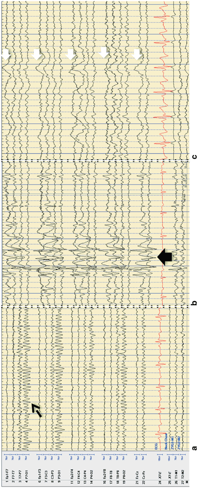
The routine EEG is safe, inexpensive, comfortable to the patient, and takes about 2 h to complete. An EEG can be repeated as often as necessary. Figure 3.1 demonstrates typical EEG changes .

Fig 3.1
a Normal EEG tracing with dashed arrow indicating alpha rhythm of 8–9 Hz posteriorly. b Abnormal EEG tracing with epileptiform abnormalities with black block arrow showing a series of spike-and-wave complexes. c Abnormal EEG tracing showing slowing of brain rhythms with a right-sided predominance (white block arrows indicating a slow wave). (Courtesy of Dr. Glen Fenton)
Electromyogram (EMG)
The EMG is the evaluation of the electrical function of individual muscle motor unit potentials at rest and during muscle contraction. The EMG is performed by inserting a recording needle electrode into the belly of a muscle . The needle tip is the recording electrode and the needle shaft is the reference electrode in a concentric needle while a monopolar needle compares the electrical signal of fibers to that of a reference electrode on the skin surface. Electrical activity from muscle fibers is recorded and amplified to appear on an oscilloscope as a tracing of voltage versus time with accompanying sound. Physicians need special training to perform and to interpret the EMG.
Abnormal motor units or individual muscle fibers demonstrate changes in duration, amplitude, and pattern of the waveform that occur during needle insertion, rest, and voluntary contraction. An EMG distinguishes normal muscle from disease due to nerve damage or muscle disease. An EMG is safe, somewhat uncomfortable to the patient, inexpensive, and requires 30–60 min. To minimize patient discomfort, the patient should receive a clear description of what will happen and frequent reassurance.
Normal Muscle
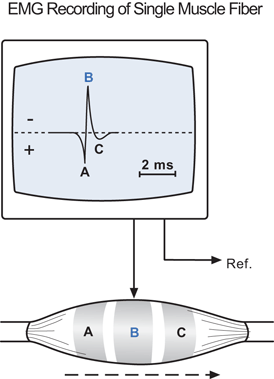
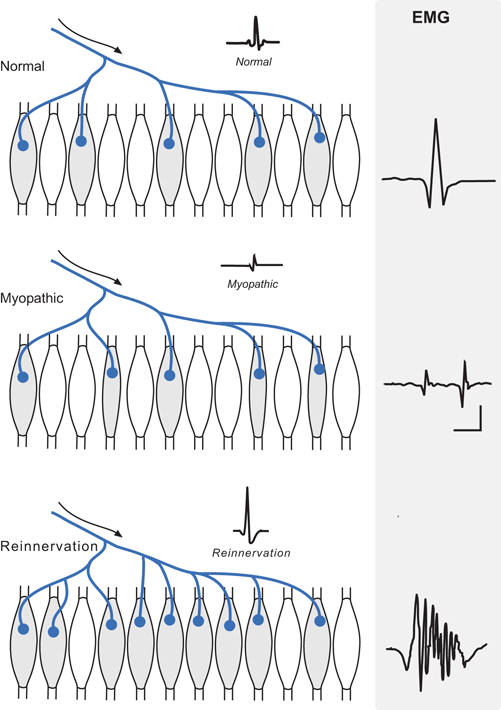
Insertion of a needle into a normal muscle injures and mechanically stimulates many muscle fibers, producing a burst of action potentials of short duration (< 300 msec) . At rest normal muscle is electrically silent as normal muscle tone is not the result of electrical contraction of muscle fibers. As an electrical impulse travels along the surface of a single muscle fiber toward the recording electrode, the impulse becomes positive (downward deflection shown on the computer screen by convention) relative to the reference electrode. As the impulse comes beneath the electrode, the waveform becomes negative (upward deflection) and then becomes slightly positive and returns to baseline as the impulse travels past the electrode (Fig. 3.2). A single muscle fiber contraction lasts about 2–4 msec and is less than 300 volt in amplitude. The firing of a single muscle fiber (called fibrillation which does not cause visible muscle movement) does not occur normally and is a sign of muscle membrane instability either from denervation or myopathy. In normal muscle, an electrical impulse travels from a spinal cord anterior horn neuron (lower motor neuron) along its axon to eventually innervate 10–1000 muscle fibers (called a motor unit). The number of muscle fibers innervated depends on the muscle with proximal limb muscles having the highest number of innervated muscle fibers. During mild voluntary muscle contraction, an entire motor unit fires almost simultaneously producing a motor unit action potential (MUAP). A typical MUAP has 3–4 excursions across the baseline (phases) and a maximum amplitude of 0.5–5 mV (Fig. 3.2) . The shape and duration of a given MUAP remain quite constant on repeat firings and generally appear different from other nearby MUAPs.
Denervated Muscle
Immediately after complete nerve transection, the muscle is paralyzed, unexcitable by nerve stimulation, and electrically silent by EMG except for insertion potentials. Beginning 2–3 weeks after a muscle loses its innervation, spontaneous individual muscle fiber contractions may appear. The EMG demonstrates fibrillations and positive sharp waves (brief monophasic positive spikes). Until the motor unit completely degenerates, spontaneous firing of the MUAP also occurs (called a fasciculation which produces a visible muscle twitch). If the nerve damage is incomplete and occurred several months earlier, the denervated muscle fiber induces adjacent motor nerves to branch or sprout and send a nerve branch to re-innervate the denervated muscle fiber called sprouting. MUAPs suggestive of sprouting are of longer duration, contain more phases, and may be of higher maximum amplitude than normal.
Myopathy
Death or dysfunction of scattered muscle fibers results in MUAPs during voluntary muscle contraction that are of shorter duration and lower amplitude than normal (Fig. 3.3). Some MUAPs may be polyphasic from loss of synchronous firing. In myositis, there may be accompanying fibrillations due to inflammatory damage to adjacent motor nerve endings .

Fig 3.2
Single MUP on EMG

Fig 3.3
EMG of motor units in diseases
In myopathies that cause myotonia (such as myotonic dystrophy), insertion of the needle produces a train of high-frequency repetitive discharges in a positive sharp waveform that diminish in frequency and amplitude over a few seconds. When heard over a speaker, myotonic discharges sound like a “dive-bomber”.
Nerve Conduction and Neuromuscular Junction Studies
Nerve conduction studies are undertaken to evaluate the functioning of motor, autonomic, and sensory nerves and neuromuscular junctions . It is possible to determine actual conduction velocities for nerves in the peripheral nervous system but conduction velocities cannot be determined in the central nervous system. In the CNS, only a nerve latency time can be obtained because the CNS nerves cannot be stimulated at various points along the nerve pathway. The test is performed by a physician with special training or by a skilled technician under a physician’s supervision. The test is safe, inexpensive, mildly uncomfortable for the patient, and takes ½–1 h.
Indications for ordering nerve studies include to (1) determine whether a neuropathy is generalized or multifocal, (2) determine whether a neuropathy is mainly from demyelination or axonal loss, (3) localize the site of a nerve conduction blockade, and (4) determine and characterize neuromuscular junction abnormalities. In the common types of distal sensorimotor peripheral neuropathy, nerve studies seldom help in establishing the etiology but can confirm historical symptoms or exam signs.
Motor Nerve Function
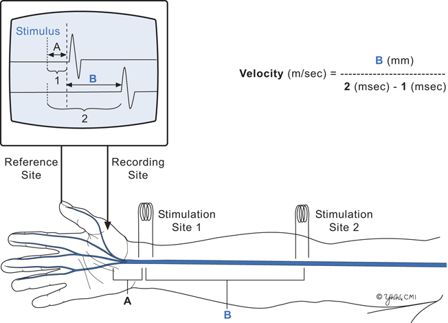
Motor nerve conduction velocity studies measure the velocity of the fastest motor nerve axons at various points along a peripheral nerve. Peripheral nerves can be stimulated to fire by application of an electrical impulse to the skin overlying the nerve. When a muscle contracts, its electrical signal can be detected by placing an electrode on the skin above the muscle belly. The muscle electrical signal is recorded, and the time from electrical stimulus to muscle contraction (latency) can be determined and displayed on an oscilloscope. A motor nerve velocity is determined as follows (Fig. 3.4). By moving the stimulating electrode along the nerve pathway, differing latencies in msec to muscle contraction are determined. By measuring the distance along the nerve pathway between two stimuli, one can divide the nerve distance in mm by the latency difference in msec to obtain the nerve velocity in meters per second. Normal motor velocity of the median and ulnar nerves is 50–60 m/sec and of the sciatic nerve is 40–50 m/sec. Slowing of the motor nerve velocity may reflect loss of myelin along the nerve (often causing slowing of velocities to 20–30 m/sec) or loss of the fastest motor nerves (lesser degree of velocity slowing). Slowing of a motor nerve may occur along the entire nerve pathway or at a localized point of nerve compression, such as the ulnar nerve at the elbow.
Sensory Nerve Function
Evaluating sensory nerve function is more difficult as the normal signals are weaker and more diffuse following an electrical stimulus because the conduction velocities of different sensory axons vary considerably. Sensory nerves may be unmyelinated and conduct at ½–2 m/sec or thinly myelinated and conduct at 10–20 m/sec. The most common sensory nerve test determines the latency time from electrical stimulation of the skin of a finger to a skin recording electrode site over the median nerve just proximal to the wrist. A delayed median nerve sensory latency suggests compression of the nerve at carpal tunnel. The test is safe, mildly expensive, somewhat uncomfortable, and takes about 30 min.
Neuromuscular Junction Function
Information about the function of the neuromuscular junction can be obtained from repetitive nerve stimulation studies. Placement of a skin recording electrode over the belly of a muscle and stimulating the motor nerve produce a compound muscle action potential (CMAP) . If the nerve stimulation is repeated, the CMAPs appear identical on the oscilloscope. In diseases of the neuromuscular junction, the amplitude of the CMAPs may decrease or increase. In myasthenia gravis and botulism , repetitive nerve stimulation produces a decremental response in the CMAP. The test is safe, inexpensive, somewhat uncomfortable, and takes about 15 min.
Sensory Evoked Potentials
Occasionally, there are indications to evaluate the integrity of central conduction along major sensory pathways (visual, auditory, and peripheral sensory system) called evoked potentials. As noted above, actual conduction velocities cannot be obtained but central modality-specific latencies can. Evoked potential tests record computer averages of the EEG that is time-locked to repeated (100–500 trials) specific sensory stimuli such as sound, light, or electrical stimulation of peripheral nerve. The computer averaging reduces background EEG electrical activity to zero while enhancing the time-locked stimulus signal. Abnormalities are characterized by a delay for the time-locked signal average to reach its destination or distortions (usually a prolongation of the waveform and loss of signal amplitude). Sensory evoked potentials are safe, inexpensive, and comfortable. The major indication is the evaluation of possible diseases that cause CNS demyelination of these sensory pathways.

Fig 3.4
Motor nerve conduction velocity diagram
Structure
Lumbar Puncture (LP) and Cerebrospinal Fluid (CSF) Examination
Five important reasons for lumbar puncture are to (1) diagnose infections of the meninges, (2) diagnose cancer involving the meninges, (3) diagnose herpes simplex encephalitis and other encephalitides, (4) diagnose a small subarachnoid hemorrhage , and (5) introduce medications into the subarachnoid space or contrast media for a myelogram. In addition, there are several diseases where examination of CSF helps make a specific diagnosis. These diseases include multiple sclerosis , Guillain–Barre syndrome, and paraneoplastic syndromes. The LP is not limited to establishing diagnoses. Antimicrobial and anticancer drugs can be delivered intrathecally into the lumbar or cisternal CSF to treat patients with some forms of infectious meningitis or meningeal carcinomatosis. The LP is safe, mildly uncomfortable, and moderately expensive depending on tests ordered and takes up to 1 h.
Stay updated, free articles. Join our Telegram channel

Full access? Get Clinical Tree


