Task
Instructions
Scoring
Date orientation
“Tell me the date?” Ask for omitted items
One point each for year, season, date, day of week, and month
5
Place orientation
“Where are you?” Ask for omitted items
One point each for state, county, town, building, and floor or room
5
Register three objects
Name three objects slowly and clearly. Ask the patient to repeat them
One point for each item correctly repeated
3
Serial sevens
Ask the patient to count backwards from 100 by 7. Stop after five answers (Or ask them to spell “world” backwards)
One point for each correct answer (or letter)
5
Recall three objects
Ask the patient to recall the objects mentioned above
One point for each item correctly remembered
3
Naming
Point to your watch and ask the patient “what is this?” Repeat with a pencil
One point for each correct answer
2
Repeating a phrase
Ask the patient to say “no ifs, ands, or buts”
One point if successful on first try
1
Verbal commands
Give the patient a plain piece of paper and say “Take this paper in your right hand, fold it in half, and put it on the floor”
One point for each correct action
3
Written commands
Show the patient a piece of paper with “CLOSE YOUR EYES” printed on it
One point if the patient’s eyes close
1
Writing
Ask the patient to write a sentence
One point if sentence has a subject, a verb, and makes sense
1
Drawing
Ask the patient to copy a pair of intersecting pentagons onto a piece of paper 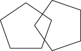
One point if the figure has ten corners and two intersecting lines
1
Scoring
A score of 24 or above is considered normal
30
Mood and affect are assessed during the mental status exam as psychiatric disease such as depression can present with memory complaints. When evaluating affect, it is important to note inappropriate tearful or jocular behavior that may not be congruous with the subject matter being discussed.
Speech and language abnormalities are divided into dysarthria and dysphasia. Dysarthria results from poor articulation—like talking with rocks in your mouth. The sentence makes sense but the sound is garbled. Abnormalities of the mouth (poor dentition) or CN IX, X, and XII dysfunction are common causes. Dysarthria does not affect the ability to read or write. Dysphasia implies dysfunction in constructing or understanding language. In expressive aphasia , the patient often speaks short truncated sentences without adjectives or adverbs but understanding is relatively preserved. Receptive aphasia usually has normal sounding speech but the content does not make sense relative to the question. In both dysphasias, there is difficulty in repeating phrases such as “No ifs ands or buts.” Dysphasia also affects the ability to read and write. Language abnormalities and apraxias are fully covered in the chapter on higher cortical function
Cranial Nerves
I. Olfaction is seldom routinely tested unless the patient has a complaint of poor taste or smell or history suggesting problems with frontal lobes or facial bones. First, ensure that there are no obstructions in the nasal passages by inspection with otoscope. Smell cannot be tested on each side separately since both sides of the nose communicate. Ask the patient to close their eyes and to identify the odor when presented and then identify the odor’s name. Common substances such as coffee grounds, unlit cigarettes, and perfumed soap are convenient to test. Use of alcohol or ammonia should not be used as those odors stimulate CN V fibers located in the anterior nose and give a false-positive test.
II. Optic nerve function is usually divided into visual acuity, visual fields, and fundoscopic exam. To test visual acuity in each eye with their glasses, one can use a Snellen eye chart or a near-vision card. Ability to read standard newsprint suggests 20/40 or better acuity. If their glasses are not available, a pinhole card (paper with pin pushed through the center) can improve their vision. If visual acuity is 20/50 or better, the problem is usually ocular and not neurologic.
Visual fields are evaluated by confrontation testing each eye separately. Standing about 4 feet away with one eye closed and the patient looking at your nose, the patient is asked to count the number of fingers (1, 2 or 5) presented in the four visual quadrants. Confrontational testing can detect a homonymous hemianopia or quadranopia but not constriction of visual fields from glaucoma.
On fundoscopic examination, carefully observe the retinal vessels for hemorrhages and exudates and then follow them into the optic disc itself. Color and size of the disc and the presence of papilledema are particularly important. Papilledema is suggested by swollen optic disc heads with the margins appearing blurred/raised.
Pupil size and the light reflex involve CN II and autonomic eye nerves. Observe the pupils in dim light with illumination from below. The pupils should be round and be within 1 mm of each other in size and constrict equally when the patient attempts to look at their nose ( accommodation). Anisocoria or unequal pupil sizes signifies dysfunction of sympathetic nerve (small pupil or miosis) or parasympathetic nerve (large pupil or mydriasis). In the light reflex, one tests a direct light reflex (the pupil constricts when a light is shined into it) and then a consensual reflex (the opposite pupil constricts when a light is shined into the other). Both pupils should constrict briskly and equally to light. Shining the light into one eye and failing to see both pupils to constrict imply ipsilateral retina or CN II dysfunction, failure of ipsilateral iris to constrict implies dysfunction of ipsilateral sympathetic nerve, and failure of contralateral pupil to constrict suggests dysfunction of contralateral sympathetic nerve.
III, IV, VI. Oculomotor, trochlear, and abducens nerves innervate the extraocular eye muscles . They are evaluated by observing the eye move correctly when the patient is asked to follow your finger in all nine directions of gaze (Fig. 2.1). Observe whether the eye movements are conjugate (move together), move the entire range, and are smooth. Presence of double vision in one gaze direction suggests dysfunction of a given nerve or eye muscle. In Fig. 2.2, a patient with a right CN VI palsy is unable to move his right eye laterally. Nystagmus can be seen in healthy people at the far end of horizontal gaze, but is an abnormal sign at rest, near mid position or is sustained.
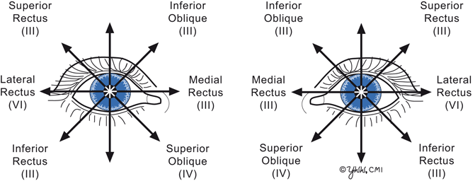
Fig. 2.1
Directions of gaze
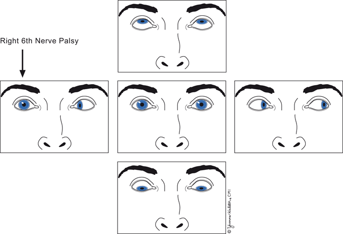
Fig. 2.2
Right 6th nerve palsy
The size of the palpebral fissure (distance between upper and lower eyelid) depends on CN III and sympathetic nerves. Marked drooping of the upper eyelid (ptosis) that interferes with vision implies CN III dysfunction or prior eye trauma. Mild ptosis without obstructing vision implies sympathetic nerve dysfunction. When mild ptosis is accompanied by ipsilateral miosis (pupillary constriction), the lesion is called Horner’s syndrome.
V. Trigeminal nerve function is tested by evaluating face sensation. Lightly touch the 3 divisions of the CN 5 with a cotton tip, your fingers or a cool tuning fork. The patient should perceive these as equal on both sides. The corneal reflex (touching the edge of the cornea over the outside of the iris with a wisp of cotton or a soft facial tissue) should produce prompt blinking of both eyes. Failure to blink in either eye suggests an afferent problem in the stimulated CN V, failure of ipsilateral eye but not contralateral eye to blink suggests dysfunction of ipsilateral CN VII, and failure of contralateral eye to blink but not ipsilateral eye suggests dysfunction of contralateral CN VII. Having the patient open her jaw and attempt to move the jaw laterally against resistance test motor fibers of CN V.
VII. Facial nerve function is evaluated by testing facial muscles. Ask the patient to open her eyes wide, close them shut tightly and pull back her lips. The muscles of facial expression, innervated by CN VII, should show equal and symmetrical movement on both sides of the face.
A lower motor neuron lesion (facial nerve or nucleus) produces weakness of both the upper and lower face. An upper motor neuron lesion (corticobulbar tract above the level of CN VII nucleus) causes weakness only of the lower face because forehead muscles receive bilateral innervation.
The chorda tympani nerve branch can be tested by determining whether the patient can detect the taste of sugar or salt placed on the anterior two-thirds of one side of the tongue.
VIII. Auditory nerve hearing evaluation is tested by masking the opposite ear with finger or sounds and determining whether the patient can hear whispers (mid sound frequencies) or rubbing fingers (higher sound frequencies) in the other ear. If there is hearing loss, the external auditory canal should be inspected with an otoscope. Vestibular nerve testing is described in the chapter on dizziness and vertigo .
IX. Glossopharyngeal nerve function is tested by asking patient to say, “aah” and observing the soft palate and uvula rising symmetrically. Deviation of the uvula and soft palate to one side indicates a lesion on the contralateral nerve. Touching the pharynx with a cotton Q-tip should elicit a gag reflex from CN IX and X.
X. Vagus nerve function is tested by listening for hoarseness in the patient’s voice. If present, vocal cord movements can be visualized by otolaryngology to confirm paralysis.
XI. Accessory nerve function is tested by shoulder shrug and head turn. Ask the patient to shrug her shoulders to her ears and then push down. Strength should be symmetric. Then ask her to turn her head to either side while you apply resistance with your entire hand on her lower jaw. Again, strength should be symmetric. Remember that the right sternocleidomastoid muscle turns the head to the left.
XII. Hypoglossal nerve function is evaluated by asking a patient to protrude the tongue straight out and moving it from side to side. Deviation of the tongue to one side with atrophy and fasciculations in that side of the tongue suggests an ipsilateral lower motor neuron lesion.
Neck: The patient should be able to smoothly flex her neck to touch her chin on the chest and rotate the head fully towards the shoulders. In meningitis, the patient cannot flex or resists flexing the neck while in cervical arthritis, there is restricted rotation of the neck, called meningismus.
Motor Examination
A complete motor examination includes the evaluation of muscle bulk, tone, strength, and gait . In addition, any involuntary movements should be noted.
Related posts:

Stay updated, free articles. Join our Telegram channel

Full access? Get Clinical Tree


