Craniopharyngiomas

Chapter 24 Craniopharyngiomas


Craniopharyngiomas are tumors of neuroepithelial origin that arise from squamous cell rests found along the path of the primitive craniopharyngeal duct. Their incidence ranges between 0.5 and 2.5 per 100,000 person years and does not vary by sex or race. Craniopharyngiomas account for 1.2% to 4.6% of all intracranial tumors (Central Brain Tumor Registry of the United States). They exhibit a bimodal distribution, first peaking during childhood (5–14 years) and later peaking in adults ranging from 50 to 74 years; they comprise 5% to 10% of pediatric brain tumors and 1% to 4% of adult brain tumors.14 Craniopharyngiomas have a growth pattern that is often in close proximity to the pituitary infundibulum, and can occur within the sella, suprasellar space, or third ventricle, frequently spanning these spaces. These tumors tend to involve a number of neural structures, including the optic nerves, internal carotid arteries (ICAs), and pituitary gland, causing a variety of symptoms. Common clinical presentations include visual dysfunction with symptoms of chiasmatic as well as postchiasmatic compression, hypothalamic dysfunction with behavioral changes ranging from alterations in eating patterns, to apathy, and even obtundation and pituitary dysfunction, often manifesting as hypopituitarism.


During the past several years, treatment algorithms have evolved that now incorporate multiple modalities. Surgical resection remains the primary treatment whenever possible. However, the suprasellar space is replete with important neurovascular structures that include the perforator arteries supplying the optic chiasm, hypothalamus, and basal ganglia; their interruption results in permanent neurologic loss of function and disability. Proven therapies with low morbidities include nonradical surgical resection followed by fractionated radiation therapy, radiosurgery, cystic lesion aspiration with implantation of Ommaya reservoir for intracavitary radioisotope, or chemotherapy instillation. More recently, surgical resection via an expanded trans-sphenoidal resection has been advocated as the most direct route to the bulk of the lesion. Although the access provided through this corridor is unparalleled, this technique remains in development because of the lack of appropriate instrumentation to allow safe dissection and the difficulty in providing a watertight closure.


The natural history of craniopharyngiomas following treatment is one of recurrence. The need for re-treatment arises when these indolent lesions that exhibit slow growth in both their solid and cystic components become symptomatic at various times during a patient’s lifetime.


Treatment Decision Making


The primary treatment of craniopharyngiomas remains surgical. The variability of the tumor extent make is imperative to tailor the surgical approach to the particular lesion. Of several different approaches described, no approach is overall preferred for the majority of the lesions. Adjuvant treatment with radiation therapy is often used; it is necessary in all cases when gross total resection is not achieved and in some tumor recurrences that appear after apparent gross total resection and are likely the result of microscopic residual disease.


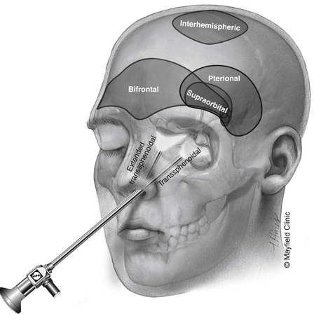
Although most patients undergo both surgical and radiation treatments, a number of questions need to be addressed on an individual level. Choice of surgical approach depends primarily on the extent of the lesion along the vertical axis (Fig. 24-1). Lesions that are purely intrasellar are preferably approached through a trans-sphenoidal approach. Microscopic or endoscopic corridors have been well described and mimic the approach to pituitary adenomas. Because intrasellar lesions are often cystic, obliteration of the tumor cavity with fat may not be indicated unless a cerebrospinal fluid (CSF) leak is manifested intraoperatively, effectively allowing for a prolonged outlet that can delay or prevent future reaccumulation of fluid within the cavity. The trans-sphenoidal route can also be used successfully in the presence of significant suprasellar extension in mostly cystic lesions. The recent development of expanded endoscopic trans-sphenoidal approaches to the sella make the resection of the cyst wall possible when vascular adherence is not a significant issue.


Lesions with significant suprasellar extension, particularly when mostly solid, are preferably approached through intracranial corridors. Although expanded endoscopic trans-sphenoidal approaches can provide effective tumor resection, they are considered alternative approaches because of the dearth of appropriate instrumentation, which can cause difficulty in safely managing intraoperative bleeding. Additionally, the dural opening immediately inferior to the suprasellar cistern has proven to be difficult to seal effectively against CSF leaks. Finally, most lesions that are primarily suprasellar arise superior to the pituitary gland, displacing it inferiorly, making the mobilization of the gland itself a necessity for access to the lesion through a trans-sphenoidal route, a surgical maneuver that can result in hypopituitarism.


Transcranial routes to the suprasellar space range from a supraorbital corridor to the pterional approach (with or without an orbital or orbitozygomatic osteotomy) to the bifrontal craniotomy. Although each one of these approaches provides a similar exposure to lesions in the suprasellar space, they differ in several important ways. The supraorbital craniotomy, which minimizes soft-tissue morbidity and creates a shorter overall incision, is limited by the size of the frontal sinus. Violation of the frontal sinus through the supraorbital approach can cause a CSF leak that is challenging to fix or a rotation periosteal flap is difficult. Additional limitations of the supraorbital craniotomy include the limited vertical exposure and difficulty with effective brain retraction.


The bifrontal subfrontal approach allows for a midline or medial exposure to the suprasellar space and can be the most effective in the presence of a post-fixed chiasm. Conversely, a prefixed chiasm is a limitation that may make access to the lesion all but impossible. Close study of preoperative imaging may give some indication to the location of the chiasm. Specifically, assessment can involve both direct visualization of the chiasm in the absence of severe distortion of normal anatomy and determination of the position of the anterior communicating artery as a surrogate for the location of the chiasm. Nonetheless, large lesions often make it difficult to ascertain the exact position of the chiasm preoperatively; in such cases, this approach can be limiting.


The pterional approach is the most versatile for accessing the majority of craniopharyngiomas. It allows for working channels in between the two optic nerves, between the optic nerve and ICA, and between the ICA and oculomotor nerve. The pterional approach is the one most often used for removal of the optic canal roof and anterior clinoid process, steps often necessary for safe mobilization of the optic nerve. The limitation of this approach relates to the location of the ipsilateral optic nerve in the direct line of sight; that is, resection of a significant part of the tumor deep to the nerve is difficult without substantial mobilization of the nerve itself.


Tumors that extend extensively into the third ventricle or infrequently arise primarily in the third ventricle pose the greatest surgical challenge, particularly when a significant cystic tumor component is absent. The two major corridors to such lesions are the translamina terminalis approach and the transventricular approach. The lamina terminalis forms the superior continuation of the optic chiasm; its fenestration allows for entry into the anterior third ventricle. It provides direct access to the anterior part of the lesion, yet is limited by the amount of retraction of the optic tracts and the anterior communicating artery necessary for visualization into the third ventricle. In large lesions, determination if a plane of safe dissection exists between the lateral walls of the tumor and hypothalamus can also be challenging. This determination is particularly difficult through a pterional approach that leads to blind dissection of the ipsilateral tumor margin.


Transventricular approaches are the most versatile to access large lesions within the third ventricle. A transfrontal or interhemispheric approach can be used to access the lateral ventricle. A transforaminal approach expanded through a subchoroidal extension provides wide access into the third ventricle and good visualization of the lateral margins of the lesion. Limitations of this approach are the significant depth at which the lesion is encountered, the thalamostriate vein that needs to be mobilized, and the potential for injury to the fornices.


Radiation therapy is often used as adjuvant therapy. However, a good indication for its use as primary treatment is the rare case of a solid third ventricular lesion with evidence of poor margins with the hypothalamus in an older patient. The radiation delivery method varies depending on the tumor’s size and characteristics. The usual limitations for radiosurgical treatment, relating to both lesion size and proximity to the optic apparatus, hold true and often lead to the need for fractionated treatment.


For primarily cystic tumors, stereotactic- or endoscopic-transventricular cyst aspiration through an implanted catheter can allow for intracystic instillation of radioactive material or chemotherapy as well as subsequent percutaneous aspirations of fluid re-accumulation.


Surgical Techniques


Trans-Sphenoidal/Expanded Trans-Sphenoidal Approach


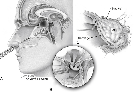
The patient is positioned supine; the head in the “sniffing” position allows for elevation above the level of the heart and drainage of bloody material inferiorly away from the surgical field. The traditional trans-sphenoidal approach through a microscopic or endoscopic approach can be used. Intrasellar tumors and mostly cystic craniopharyngiomas even with a significant suprasellar extension are excellent candidates for this approach (Fig. 24-2).


The expanded trans-sphenoidal approach requires significant bony removal superiorly past the tuberculum. Importantly, at the beginning of an expanded trans-sphenoidal approach, consideration should be given to raising a nasoseptal mucosal flap, which will be used during closure.20 Visualization of the superior aspect of the lesion and its relationship to the undersurface of the optic chiasm is feasible with debulking of the tumor. The perforating vessels and the plane with the optic apparatus need to be sharply dissected under direct vision. However, present endoscopic instrumentation creates an obstacle to effective dissection in many patients.


Closure of a traditional or expanded trans-sphenoidal approach has important considerations. Intrasellar lesion resection can be associated with CSF leaks. Obliteration of the sella with abdominal fat grafting is our preferred reconstruction technique; it is reinforced by bone or cartilage obtained during the exposure and by tissue sealant onlay over the entire reconstruction. In the absence of a CSF leak, one may consider leaving the sella open to delay or obviate the re-accumulation of fluid within the cyst. Caution should be used in making this decision depending on the amount of arachnoid descent and herniation within the sella because a delayed CSF leak may ensue.


Although a watertight closure of expanded trans-sphenoidal approaches remains elusive, significant steps have been made during the past several years. Simple fat obliteration proves to be inadequate reconstruction. Multilayer reconstructions and the use of vascularized pedicled mucosal flaps have been promising as an effective reconstruction that can prevent leaks. Use of specialized clips that attempt direct dural reapproximation is an obvious improvement, yet technically very challenging. Deployment of inflatable balloons within the sphenoid sinus can augment the reconstruction. Lumbar subarachnoid drains for temporary CSF diversion offer mechanical advantages but must be balanced by their risk of potential complications, compression of neurovascular structures for the former, and development of pneumocephalus for the latter.


Bifrontal Craniotomy


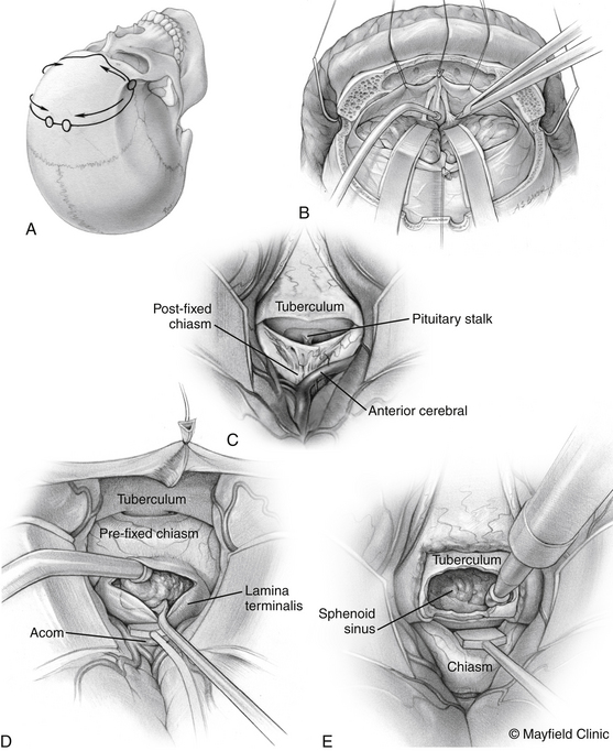
The patient is positioned supine with the head slightly extended in three-point pin fixation in the Mayfield clamp. Frameless stereotactic guidance is a useful adjunct for accurate localization of the lesion. A bifrontal incision is marked behind the hairline. Ensuring that the lateral limits are close to the zygomas bilaterally then limits the pressure on the skin flap and avoids flap ischemia. Burr holes are placed on either side of the superior sagittal sinus, approximately 5 cm above the nasion and laterally superior to the keyhole, just lateral to the superior insertion of the temporalis muscle, which is elevated in a limited fashion. The inferior bony cut is preferably made just superior to the frontal sinus; however, transgression of the sinus can be fixed easily with the use of a rotational pericranial flap. The dura is opened in a horizontal incision inferiorly and the superior sagittal sinus is divided with medium vascular clips. The falx is incised and the anterior interhemispheric space is explored (Fig. 24-3).


Once the lesion is localized, its relationship to the optic nerves and chiasm is assessed. A prefixed location of the chiasm is a limitation of this approach. In the absence of significant extension into the third ventricle, drilling the tuberculum often allows for increased exposure subchiasmatically. Although exposure is potentially adequate for resection of the lesion, this is a limited corridor for large lesions. Reconstruction of the skull base is crucial for a good outcome.


The anterior interhemispheric approach is perhaps the optimal approach for lesions with extension into the third ventricle. After exposing the chiasm, the surgeon identifies the lamina terminalis, dissects and gently retracts the anterior communicating artery superiorly, and a fenestration into the third ventricle. A midline fenestration at the thinnest area, as far superior to the chiasm as feasible, is most desirable. Correct identification of the optic nerves and optic tracts bilaterally is the safest way to avoid injury to the visual system.


Dissection of the tumor from the third ventricle can be safely achieved through the fenestration of the lamina terminalis. The principles that ensure safety and improved outcome are careful identification of the visual apparatus and careful dissection of the lateral margin of the tumor from the hypothalamus. In cases where the plane between the tumor and the lateral wall of third ventricle is not directly visible or absent, one needs to exercise caution to avoid hypothalamic dysfunction.


Pterional Craniotomy


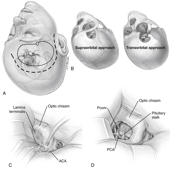
The traditional frontotemporal or pterional craniotomy is perhaps the most widely used approach for the treatment of craniopharyngiomas. Reduction of the sphenoid wing is a standard part of this approach because it improves exposure superiorly. Once the basal cisterns are opened, there are two major corridors for lesion resection: the subchiasmatic corridor between the two optic nerves and the opticocarotid corridor between the lateral aspect of the ipsilateral optic nerve and ICA (Fig. 24-4).


This approach is familiar to most neurosurgeons, yet its relative limitation is that the lesion lies posterior to the ipsilateral optic nerve, which often is displaced anteriorly and superiorly as it is splayed over the tumor surface. Manipulation of the ipsilateral optic nerve is a maneuver that endangers visual function. Therefore, one needs to resist the temptation to mobilize the optic nerve for enhanced tumor exposure. Section of the falciform ligament and fenestration of the optic canal, and less often anterior clinoidectomy, are surgical adjuncts that minimize the risk to the ipsilateral vision.


The pterional approach allows for direct visualization of the microvasculature surrounding the lesion in the suprasellar cistern. Careful dissection of perforating vessels from the mesial wall of the ICA and the anterior cerebral artery prevents vascular injury to the anterior perforating substance, basal ganglia, hypothalamus, and visual system. The pterional approach is also a versatile one. An orbital osteotomy allows for wide access and improvement in the superior aspect of the surgical exposure. Dissection along the ipsilateral optic nerve and chiasm allows for fenestration of the lamina terminalis and access to the part of the lesion extending into the third ventricle. Care should be taken to correctly identify the location of the lamina terminalis. The 15- to 45-degree rotation off the vertical axis can disorient the surgeon with respect to midline, particularly at depth. Confirmation of midline anatomically is crucial with respect to the anterior communicating artery as well as with the use of frameless stereotactic guidance. Off-midline fenestration may result in both poor access to the lesion and injury to the visual system and hypothalamus.


The supraorbital craniotomy and its transorbital variant provide a similar degree of exposure with somewhat limited soft tissue dissection (Fig. 24-4B). The temporalis muscle is mobilized to a much lesser degree. With the incision is linear within the eyebrow, there is little need for subcutaneous dissection. The bone flap, which averages 3 × 2 cm, provides adequate access to most lesions in the parasellar space. Care should be taken to avoid injury to the frontalis branch of the facial nerve along the lateral aspect of the incision. The use of frameless stereotactic guidance is recommended to prevent violation of the lateral recess of the frontal sinus. Reconstruction of the sinus from this approach is limited and may result in a transnasal CSF leak that can prove difficult to seal. In our practice, an extensive lateral recess of the frontal sinus is a contraindication for this approach.

< div class='tao-gold-member'>

Stay updated, free articles. Join our Telegram channel

Jul 16, 2016 | Posted by in NEUROSURGERY | Comments Off on Craniopharyngiomas

Full access? Get Clinical Tree

Get Clinical Tree app for offline access