Chapter 135 Decompressive Craniectomy for Traumatic Brain Injury
Introduction
Traumatic brain injury (TBI) is a significant cause of death and disability, accounting for an estimated 294,000 hospitalizations and 52,000 deaths annually in the United States alone according to 2006 Centers for Disease Control and Prevention data.1 Although neurosurgeons play an integral role in the management of TBI, much controversy exists regarding the use of surgical therapies, especially decompressive craniectomy. Current guidelines for the treatment of severe TBI recommend decompressive craniectomy as a salvage therapy for medically refractory intracranial hypertension,2 but little class I evidence exists comparing decompressive craniectomy to nonsurgical management for the treatment of TBI in adults.3,4 Many reviews have been written discussing the relevant data for and against the use of decompressive craniectomy for TBI, with the overwhelming conclusion being that randomized controlled trials are needed to resolve these disputes. The goal of this chapter is to summarize the data pertaining to the use decompressive craniectomy for TBI and to discuss the various issues associated with this procedure, including outcomes, techniques, and complications.
Historical perspective
Evidence of surgical decompression performed to treat TBI goes as far back as Ancient Egypt and Ancient Greece, and the indications for its use included TBI, epilepsy, headache, and mental illness.5 The concept of using surgical decompression to treat elevated intracranial pressure (ICP) was introduced to modern neurosurgery by Kocher6 and Cushing7,8 at the beginning of the 20th century. Discussion of surgical decompression for TBI in the literature did not become widespread, however, until the late 1960s and 1970s after a series of studies examining surgical decompression for various TBI-related entities. At that time, the benefit of surgical decompression was not clear. Jamieson and Yelland9 reported encouraging results for decompression of traumatic epidural hematomas, with a mortality rate of just 16% in a series of 167 patients. Surgical techniques used in their series ranged from subtemporal and suboccipital decompressions to “local” craniectomies, trephines, and osteoplastic craniotomies. Subsequent review of surgical management of traumatic subdural hematomas by the same authors, however, showed worse outcomes. In a series of 555 surgically managed subdural hematomas, 317 of the patients underwent a decompressive craniectomy, with an associated 43% mortality rate.10 Similarly, surgically managed intraparenchymal hematomas were associated with a 24% mortality rate,11 although the outcomes associated with various surgical techniques were not clearly described.
In the late 1960s and early 1970s, several groups began to study specific decompressive techniques for the treatment of severe TBI. Kjellberg et al.12 reported using a bifrontal decompressive craniectomy with duraplasty for intractable cerebral edema in 73 patients, 50 of whom had sustained TBI. They reported an 18% survival rate (22% among TBI patients), as well as an additional 16 patients who showed neurological improvement postoperatively but died as a result of other medical complications. Similarly, Venes et al13 reported 13 TBI patients with intractable cerebral edema treated with wide decompressive bifrontal craniectomies. Although the mortality rate was only 31%, only one patient (a 2½-year-old child with moderately severe TBI) returned to normal neurological function postoperatively with the exception of a seizure disorder. A wide decompressive hemicraniectomy (DHC) with durotomy was described by Ransohoff et al.14 for the treatment of subdural hematoma. They reported a 40% survival rate, and 28% of patients returning to normal activities. These outcomes were much improved over an 85% mortality rate among TBI patients treated with burr holes or routine craniectomies.14 A follow-up study by the same group, however, showed much worse outcomes for traumatic subdural hematomas treated with decompressive craniectomy, with only a 10% survival rate.15 The authors attributed this difference to the presence of concomitant brain stem or subcortical injury. Britt et al16 reported a cohort of 42 patients who underwent decompression of an acute traumatic subdural hematoma, 34 of whom had their bone flaps left off because of cerebral edema. Although Britt et al. did not separate outcomes based on craniectomy or craniotomy, they did report an overall 55% mortality rate (only 36% mortality within 30 days of decompression) and stated that their results led them to standardize the use of a large DHC for the treatment of traumatic subdural hematomas. Because poor outcomes from subdural and epidural hematomas were partially attributed to underlying contusions, Yamaura et al.17 studied the use of DHC for severe traumatic contusions. Twenty-four percent of their patients were converted to bilateral hemicraniectomies if the “unilateral opening did not seem sufficient in its decompressive effect.” They reported an overall 36% mortality rate and advocated for the use of bilateral hemicraniectomies. In radiographic follow-up of patients who had undergone hemicraniectomy, Morantz et al.18 confirmed the benefits of DHC on the resolution of midline shift and removal of hematomas. In addition to functional outcomes, Hase et al.19 examined the effect of decompressive surgery on the elasticity (change in pressure divided by change in volume) of the brain and found that decompression decreases elasticity, thus reducing pressure variations with changes in intracerebral volume.
In an attempt to further understand the pathophysiology underlying posttraumatic cerebral edema and the role of surgical decompression, several groups have developed experimental models. Moody et al.20 studied a model of TBI using epidural balloon compression in dogs. Ten dogs received no surgical decompression, and all died within 12 hours of injury. Autopsies revealed pontine hemorrhages. A second group of dogs underwent a DHC. Although none of the surgically treated dogs regained consciousness, decompression resulted in less pontine injury. However, the hemicraniectomy also resulted in hemorrhagic infarction, necrosis, and edema at the site of the craniectomy. Cooper et al.21 studied surgical decompression in a dog model of cold-injury-induced cerebral edema. Hemicraniectomy lowered ICP but resulted in significantly greater cerebral edema. This effect was attributed to possible reduction in the interstitial pressure within the brain after decompression, resulting in a greater hydrostatic pressure gradient between the intravascular and interstitial spaces.
Overall, these studies demonstrate an increasing interest in the utility of decompressive craniectomy for TBI and provide evidence for a possible benefit regarding mortality rate after surgical decompression in severe TBI but questioned whether the morbidity and quality of life attained are justifiable. Importantly, the complex nature of TBI was recognized, and many authors acknowledged the need for a systems-based approach to the management of TBI that encompasses more than just the surgical aspects of management.22 In addition, the Glasgow Coma Scale (GCS)23 and the Glasgow Outcome Scale (GOS)24 were developed, allowing for more uniform characterization and generalization of TBI patients across centers. For the next several decades, however, few studies were published regarding the use of decompressive craniectomy in TBI.
Current Evidence
More recently, there has been renewed interested in defining the use and benefits of decompressive craniectomy, with an ever increasing number of publications on the topic over the past 20 years. Gower et al.25 began to reexplore decompressive craniectomy by reporting 10 patients treated with salvage subtemporal decompression among a series of 115 severe TBI patients. They demonstrated a mortality rate of 40% among decompressive craniectomy patients compared to 82% mortality among patients treated medically (with pentobarbital-induced coma). They also showed a 34% reduction in ICP after decompressive craniectomy. Gaab et al.26 applied both unilateral and bilateral frontoparietal temporal craniectomies and dural enlargement in patients with medically refractory cerebral hypertension and showed only a 13.5% mortality rate, with 78% achieving a GOS score of 4 or 5.
Polin et al.27 compared 35 patients with malignant posttraumatic cerebral edema treated with bifrontal decompressive craniectomy to matched controls from the Traumatic Coma Data Bank. They reported 23% mortality and 37% good outcomes (GOS score 4 or 5), a significant improvement compared to matched controls. They also demonstrated significantly lower postoperative ICP compared to controls who did not have surgery. In addition, Polin et al. identified young age and early timing of decompressive craniectomy (within 48 hr of injury) as possible favorable prognostic factors. Kleist-Welch Guerra et al.28 likewise demonstrated promising results in a prospective study of 57 patients with severe TBI and medically refractory cerebral edema, 31 of whom were treated with a unilateral craniectomy and the remainder with bilateral craniectomies. Kleist-Welch Guerra et al. reported 19% mortality and 58% favorable outcomes and advocated for the use of decompressive craniectomy before barbiturate-induced coma. In a retrospective review of DHC for severe TBI, Münch et al.29 reported higher mortality (52%) but similar favorable outcomes (41%). Similarly to Polin et al., they identified young age and early DHC as favorable prognostic factors. They also demonstrated various improvements in computed tomography (CT) characteristics after DHC, including decreased midline shift and increased visibility of the mesencephalic cisterns. These studies prompted a series of investigations into the role of decompressive craniectomy in the treatment of TBI, including indications, techniques, and prognostic factors,30–55 which culminated in a recent randomized controlled trial of surgical decompression for TBI56 as well as multiple ongoing randomized trials.
Indications and Timing
The most common indication for decompressive craniectomy in the setting of TBI has been salvage therapy for medically refractory cerebral hypertension.28,30,41,48,51,53,56,57 In this setting, ICP monitoring is utilized to guide medical management. Typical medical therapy protocols include a combination of head-of-bed elevation, cerebrospinal fluid (CSF) drainage, sedation, hyperventilation, paralysis, hyperosmolar therapy, and barbiturate-induced coma.2 Several groups, however, have also employed decompressive craniectomy at the time of initial hematoma evacuation based on the intraoperative finding of cerebral swelling.29,31,33,40,43,44,46,47,58–61 Both indications have been supported by TBI management guidelines,2,3 but one of the chief controversies in the use of decompressive craniectomy for TBI is the most appropriate timing of decompression after injury. Several studies have compared the use of early (typically within 24 hr of injury and in conjunction with hematoma evacuation) to late (more than 24 to 48 hr after injury, typically to treat medically refractory cerebral hypertension). Patients within the early and late groups, of course, are distinct populations and cannot be generalized to one another. As such, comparisons of decompressive craniectomy for each group have had mixed results, with some studies reporting superior outcomes after early decompressive craniectomy,29,47,59 some reporting worse outcomes,44 and others reporting no difference.30,60 Coplin et al.33 sought to evaluate the benefit of early decompressive craniectomy at the time of initial hematoma evacuation by comparing TBI patients with traumatic mass lesions who underwent either craniotomy or craniectomy. They found no significant differences between the two groups, despite worse injuries in the craniectomy group. Similarly, Woertgen et al.54 compared craniotomy to craniectomy for treatment of acute traumatic subdural hematoma and also found no significant difference in outcomes. As Coplin et al. also reported, the craniectomy patients in the Woertgen et al. study were found to have worse injuries intraoperatively, prompting surgeons to leave off the bone flap. The fact that both studies found similar outcomes despite worse injuries in the craniectomy groups suggests that early decompressive craniectomy, or possibly even prophylactic decompressive craniectomy, at the time of initial hematoma evacuation may in fact provide benefit.
Prognostic Factors
Several studies have examined various potential prognostic factors in an attempt to better define the patient population that will benefit most from decompressive craniectomy. The two most common, and most easily obtained, factors studied have been age and preoperative GCS score. Early studies excluded older patients, with age cut-offs as low as 30 years,28 but several studies ultimately evaluated age as a prognostic factor. Several studies have reported a correlation between age and outcomes,27,29,32,43–46,48,54,62 which is not surprising, given that age has been shown to correlate with outcome after TBI in general.63 Other decompressive craniectomy studies, however, have found no correlation.30,61 De Bonis et al.64 recently summarized the decompressive craniectomy literature with regard to age as a prognostic factor, citing studies that showed a correlation with age as well as studies that did not, and concluded that there are no strong data to support an effect of age on outcome and that the age cut-offs reported in many studies are arbitrary. At our institution, we typically do not include age in making the decision whether to proceed with decompressive craniectomy. However, on the basis of our own data suggesting that old age does correlate with poor outcomes after decompressive craniectomy for TBI, we do temper our expectations of outcomes in older patients.
GCS score is, of course, another commonly used prognostic factor in TBI,65,66 and several decompressive craniectomy studies have shown a positive correlation between preoperative GCS score and GOS score.26,32,45,52,61,62 The correlation of GCS score with outcome is likely complicated, with evidence that the motor score alone may be more prognostic than the total GCS score.67 In addition, confounding factors such as the use of alcohol and other drugs as well as the timing of any acute changes in GCS score must be considered to accurately interpret a preoperative GCS score. Response of ICP to decompression has also been shown to be a possible postoperative prognostic factor, with patients who continue to have high ICP after decompressive craniectomy being more likely to have poor functional outcomes.27,62
Preoperative Evaluation
Current guidelines recommend decompressive craniectomy as a salvage therapy for medically refractory elevated ICP in the setting of TBI.2 In this situation, patients typically already have undergone cranial imaging and ICP monitoring. Imaging studies should be reviewed to determine the most appropriate technique and laterality for decompression. Skull fractures should be identified to anticipate potential bleeding sources and to avoid cautery injury to the exposed brain tissue during the soft-tissue dissection. Because cervical spine injuries are also often associated with TBI, it is important to determine the stability of the cervical spine to avoid any cervical injuries during positioning in the operating room. Decompressive craniectomy can also be used as a prophylactic measure during emergent evacuation of mass lesions if the development of elevated ICP is deemed likely based on computed tomographic scan findings or the intraoperative appearance of the brain.31,58 When performed correctly, decompressive craniectomy can reduce ICP, increase cerebral blood flow and oxygenation, and reduce therapeutic intensity levels,30,37,53,57,59,68 potentially preventing cerebral herniation and death.
Decompressive craniectomy is most often performed in the setting of impending life-threatening cerebral herniation. It is therefore important that family members as well as the treating surgeons and physicians be aware of the patient’s dire prognosis to avoid unrealistic expectations. Decompressive craniectomy is a well-established treatment for elevated ICP, but it is unable to reverse most preexisting cerebral injuries resulting from the initial insult. This is especially true for older patients and for patients with persistently low GCS scores after the time of initial injury, who may be at greatest risk for a severely disabled or persistent vegetative state outcome. Thus, whenever possible, a realistic assessment of prognosis and potential for recovery in a specific case should be discussed prior to performing decompressive craniectomy.69 In emergent cases, however, this discussion is not always possible prior to surgery.
Technique
Several techniques have been employed for surgical decompression after TBI, but two main techniques are currently used for the treatment of medically refractory cerebral edema: DHC and the bifrontal (Kjellberg) craniectomy.70,71 To date, no study has directly compared the efficacy of these techniques, but surgeons in the majority of studies cited herein have used one or both techniques. Surgeon preference is the most common reason for choosing one over the other, although the DHC bone flap can used for traumatic hematoma evacuations, giving the surgeon the option of replacing the bone flap or leaving it off if the brain appears swollen. For each technique, the head is typically placed on a foam or rubber donut and not pinned. Rigid fixation with pins is generally not recommended in the setting of trauma, unless it is certain that there are no skull fractures. It should be noted that skull fractures parallel to the plane of CT imaging can be missed. Cervical spine precautions should also be followed if the cervical spine has not yet been cleared. Care should be taken not to compress the jugular veins, whether with tight cervical collar placement, over-rotation of the head, or placement of central venous lines on the side of the injury (if at all avoidable), as these can all further increase ICP.
Decompressive Hemicraniectomy
For unilateral DHC,70,71 the patient is placed supine with a small rolled towel underneath the ipsilateral shoulder and the head turned toward the contralateral side. Prior to prepping and draping the patient, the midline should be clearly marked and then draped outside the surgical field. Accidental transgression of the superior sagittal sinus during the craniectomy can have devastating consequences, especially in a trauma situation, when patients are often coagulopathic. Marking the midline can prevent such complications. We place the towel on the ipsilateral side of the midline to provide additional protection against crossing it.
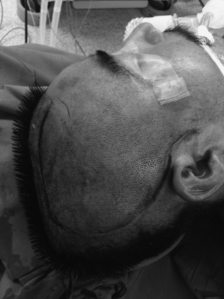
Once the site is prepped and draped, a large reverse question mark incision is made starting at the level of the zygoma and curving posteriorly above the ear, over the parieto-occipital region, then superiorly and anteriorly, approximately 2 cm lateral to the midline, and stopping just behind the hairline (Fig. 135-1). The posterior extent of the incision should be more than 15 cm behind the keyhole to allow for an adequate craniectomy flap. If possible, care should be taken to protect the superficial temporal artery to preserve blood supply to the skin flap. The incision should be extended through the subcutaneous tissue, including the temporalis muscle, down to the cranium. The resultant myocutaneous flap can then be reflected anteriorly and fixed with scalp hooks (Fig. 135-2). The temporalis dissection should be carried down to the zygoma to adequately expose the temporal bone and maximize the temporal decompression.
< div class='tao-gold-member'>
Related posts:
 Chemotherapy for Brain Tumors
Current Surgical Management of High-Grade Gliomas
Endoscopic Endonasal Approach for Craniopharyngiomas
Revascularization Techniques in Pediatric Cerebrovascular Disorders
Surgical Management of Parasagittal and Convexity Meningiomas
Surgical Management of Major Skull Defects and Potential Complications
Chemotherapy for Brain Tumors
Current Surgical Management of High-Grade Gliomas
Endoscopic Endonasal Approach for Craniopharyngiomas
Revascularization Techniques in Pediatric Cerebrovascular Disorders
Surgical Management of Parasagittal and Convexity Meningiomas
Surgical Management of Major Skull Defects and Potential Complications
Stay updated, free articles. Join our Telegram channel

Full access? Get Clinical Tree


