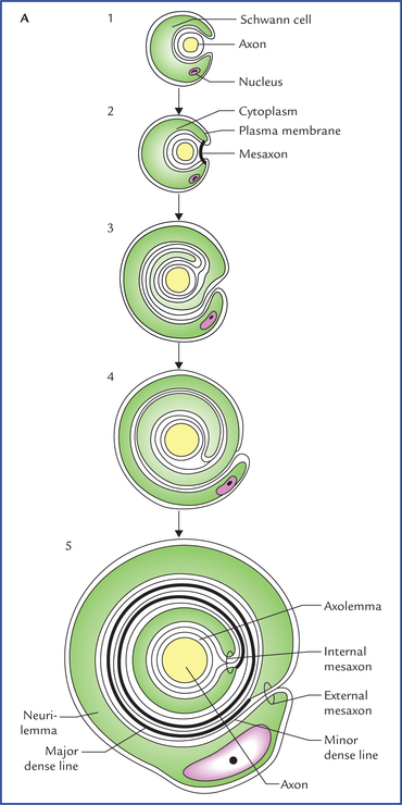
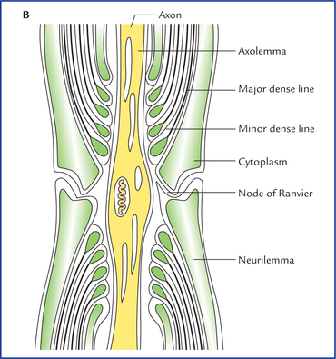
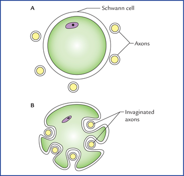
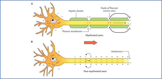
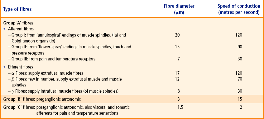
3 The peripheral nerve lesions are common in clinical practice and can be caused by a wide variety of diseases like trauma, neoplasms, infection, metabolic diseases (diabetes) and chemical toxins such as lead. The myelination begins near the origin of the axon and ends just before its terminal branches. Fig. 3.1 Myelination. (A) Stages in the formation of myelin sheath. The axon invaginates the cytoplasm of the Schwann cell and becomes suspended by a mesaxon (1 and 2). The mesaxon elongates and bound around the axon spirally (3, 4 and 5). The axon invaginates the side of a Schwann cell, as a result the plasma membrane of Schwann cell forms a mesaxon, which suspends the axon within the Schwann cell. The layer of plasma membrane immediately around the axon is continuous with the remainder of the plasma membrane through a double layered mesaxon (Fig. 3.1A). • Provides support to the nerve fibres. • Aids in conduction of the nerve impulses. • Insulates an axon from the extracellular environment. • Responsible for the colour of the white matter of the brain and spinal cord. The non-myelinated fibres are also surrounded by Schwann cells (Fig. 3.2). Several axons become longitudinally invagi-nated into the cytoplasm of a Schwann cell so that each fibre is embedded in a groove in the Schwann cell cytoplasm. The Schwann cell plasma membrane fuses along the opening of the groove, thus effectively sealing the nerve fibre within an extracellular compartment. As many as 15 or more axons may share a single Schwann cell. Fig. 3.1 (B) The longitudinal section of the myelinated nerve showing fine structure of the node of Ranvier. Fig. 3.2 Relationship of several non-myelinated axons to a Schwann cell. (A) Axons lying near the plasma membrane of a Schwann cell. (B) The axons are longitudinally invaginated into the cytoplasm of Schwann cell. Like all the cells, the resting (unstimulated) neuron maintains an ionic gradient across its plasma membrane, thereby creating an electrical potential called resting membrane potential. Thus, in resting neuron its plasma membrane remains polarized. The excitability (a fundamental property of neurons) involves a change in membrane permeability in response to appropriate stimuli so that the ionic gradient across the plasma membrane is reversed and the plasma membrane becomes depolarized. A wave of depolarization known as action potential then spreads along the plasma membrane. This is followed by the process of repolarization in which membrane rapidly re-establishes its resting potential. The speed of conduction of the action potential, along an axon depends on the myelination of the axon (Fig. 3.3). The action potentials are conducted more rapidly in myelinated than in non-myelinated axons. In non-myelinated fibres, the action potential passes continuously along the axolemma, progressively exciting neighbouring areas of membrane. In myelinated fibres, the myelin sheath serves as an insulator. Consequently a myelinated nerve fibre can be stimulated only at the nodes of Ranvier, where the axon is naked and the ions can pass freely through the plasma membrane between the extracellular fluid and the axo-plasm. Therefore, in these fibres the action potential jumps from one node to the next. The action potential at one node sets up a current in the surrounding tissue fluid, which quickly produces depolarization at the next node. The action potential conduction in a myelinated fibre is like a grasshopper jumping, whereas action potential conduction in a non-myelinated fibre is like a grasshopper walking. The action potential will naturally move more rapidly by jumping. Fig. 3.3 The conduction of action potential along an axon. (A) In myelinated axon the action potential is conducted from one node of Ranvier to another (saltatory conduction). (B) In non-myelinated axon the action potential is conducted along the entire length of the axon. The smaller sensory fibres have slower conduction rate (Table 3.1). According to the axonal diameter (including myelin sheath if present) and speed of conduction, the peripheral nerve fibres are classified into three main groups: A, B and C. • Type A fibres are large diameter, myelinated axons and therefore conduct action potentials at a great speed (15 – 120 m/sec). Motor neurons supplying skeletal muscles and most sensory neurons have type A fibres. Consequently, rapid response to external stimuli is possible as there is rapid input of sensory information to CNS on one hand and rapid output of action potential to skeletal muscle on the other hand. • Type B fibres are medium-diameter, myelinated axons and conduct action potentials at a slow speed (3-15 m/sec). • Type C fibres are small-diameter non-myelinated axons that conduct action potentials at a very slow speed (2 m/sec or less). The group A fibres are further classified into somatic sensory (I, II, III) and motor (a, (3, 7) subgroups. Table 3.1 shows the types of nerve fibres and their maximum diameters and conduction rates.
Peripheral Nerves and Ganglia
Nerve Fibres
Myelinated and Non-myelinated Nerve Fibres
Myelination (formation of myelin)
Myelination of the peripheral nerve fibres (Fig. 3.1)
Functions of the myelin sheath
Conduction of Action Potential along an Axon
Classification of Peripheral Nerve Fibres
Related posts:

Stay updated, free articles. Join our Telegram channel

Full access? Get Clinical Tree


