Fig. 11.1
Primary, secondary, and tertiary multimodal association cortices, and paralimbic regions

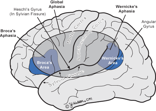
Fig. 11.2
Location of brain lesions causing different types of aphasia (left hemisphere)
Tertiary association cortex is a particularly important node for complex behaviors. While secondary association cortex receives projections primarily from its associated primary sensory cortex, the tertiary association cortex is multimodal and receives projections from multiple sensory regions. These tertiary regions include the superior and inferior parietal areas and prefrontal cortex, and they are important for complex perception and higher order abilities, such as language , complex spatial skills , and executive functions (e.g., problem solving, planning).
The limbic system includes the limbic lobe (subcallosal area, cingulate gyrus, parahippocampus, uncus, and hippocampal formation), many nuclei of the nucleus accumbens, the hypothalamus, mammillary bodies, the amygdala, and the cingulate gyrus (Fig. 11.1). The major arterial supply comes from the anterior and posterior cerebral arteries and anterior choroidal artery. The two major functions of the limbic system are emotion and memory, which is heavily dependent on circuits that include the hippocampus.
The Influence of Hemispheric Asymmetries
There is strong evidence that the left hemisphere of the brain is dominant for language, especially in right-handers. About 99 % of right-handers and 70 % of left-handers are left hemisphere dominant for language . Therefore, handedness is important information to obtain from patients in order to guide the focus of the examination. These findings show that the left and right hemispheres are often important for different higher order functions, such as language. However, it is also common for a complex task to be impaired after damage in tertiary association cortex of the left or right hemisphere even in right-handers, but the reason for the deficit is different (See Table 11.1). For example, deficits can be seen after right hemisphere damage on language tasks, such as speaking. However, expressive language abnormalities after left hemisphere damage are associated with impaired language (e.g., impaired word finding) and after right hemisphere damage are associated with impairment in the melody or prosody of speech. Table 11.1 summarizes the most important hemispheric differences in higher order cognitive functions.
Table 11.1
Deficits after damage to particular tertiary association areas in the left or right hemisphere
Left hemisphere functions | Right hemisphere functions |
|---|---|
Frontal, parietal, and temporal lobes | |
Aphasia: auditory comprehension, aspeech, areading, awriting | aSpeech (prosody), areading and awriting due to perceptual/spatial deficits |
Parietal lobe | |
Limb apraxia: deficit in what common objects are used for or deficit in the spatial and temporal characteristics of a gesture | Dressing apraxia: difficulty dressing for spatial reasons, such as inability to identify top or bottom, left or right of garment |
aConstructional apraxia: minor impairment copying or drawing of geometric forms or three-dimensional constructions due to errors of detail | aConstructional apraxia: more severe impairment copying or drawing geometric forms or three-dimensional constructions due to errors of spatial relationships (Gestalt of design); see Fig. 11.3 for examples |
Left hemineglect: inattention to the left hemispace and/or left side of body despite intact visual fields, somatosensory functions, and motor skills; see Fig. 11.4 for visual neglect | |
Medial temporal lobe-memory | |
Verbal learning and memory | Nonverbal visual learning and memory |
Frontal lobes: dysexecutive syndrome | |
Initiation of verbal information, such as rapidly naming words beginning with a particular letter | Initiation of nonverbal information, such as rapidly drawing unique designs |
Judgment, sense of purpose, planning, problem solving, cognitive flexibility, simultaneous processing, response inhibition, mental tracking | |

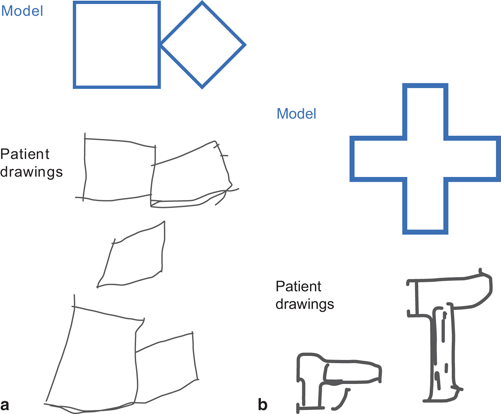
Fig. 11.3
Drawings copied by a patient with right parietal damage to illustrate the fact that a the patient could draw the two parts of the design, but could not accurately capture the spatial relationship between the two figures and b could not accurately copy a cross even with several attempts

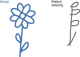
Fig. 11.4
Illustration of hemispatial neglect in a patient without visual field cut. The left side of the drawing is omitted reflecting hemispatial neglect, and the right side of the drawing illustrates visuospatial deficits
Neurobehavioral Syndromes
Aphasia
Language impairment or aphasia is seen most frequently with damage to the lateral surface of the left hemisphere. There are several types of aphasia that are differentiated by their patterns of deficits in expressive language, auditory comprehension, and naming (see Table 11.2) and the area of brain damage. Fig. 11.2 depicts the location for Broca’s, Wernicke’s, Global, and anomic aphasia, the four most important and most frequent types of aphasia.
Table 11.2
Aphasia types, descriptions, and lesion location in left hemisphere
Type | Spontaneous speech | Auditory comprehension | Repetition | Area of damage |
|---|---|---|---|---|
Broca’s, Expressive, Nonfluent | Nonfluent | Intact | Impaired | Posterior inferior frontal |
Wernicke’s, Receptive, Fluent | Fluent | Impaired | Impaired | Superior temporal gyrus |
Global, Expressive & Receptive | Nonfluent | Impaired | Impaired | Posterior frontal and temporal |
Anomic, word finding problems | Nonfluent due to word finding problems | Intact | Intact | Not localizing |
Broca’s aphasia is known as nonfluent or expressive aphasia. It is characterized by nonfluent expressive language that is agrammatic or telegraphic, such that language is dominated by subjects and verbs but not modifiers or conjunctions. Expressive language is hesitant in mild cases and minimal in more severe cases with relatively intact auditory comprehension. Associated hemiparesis is common given the proximity of Broca’s area to the motor cortex, and because these patients’ auditory comprehension is intact, they usually keep trying to self-correct errors with little success. They also can become quite frustrated with their inability to speak normally despite repeated tries. While damage to Broca’s area in the inferior third frontal convolution of the left hemisphere (Brodmann Area 44) is emphasized, neuroimaging suggests that more widespread suprasylvian damage back to the inferior parietal cortex is more common for Broca’s aphasia .
Wernicke’s aphasia is known as fluent aphasia or receptive aphasia. It is characterized by severely impaired auditory comprehension with fluent expressive language that is often not appropriate for the situation due to comprehension deficits. Paraphasic errors are common, and these patients are sometimes described as having a jargon aphasia when their expressive language is dominated by phonemic paraphasic errors (‘spoot’ for spoon), semantic paraphasic errors (‘fork’ for spoon), and neologisms (output that is not real words). Because of severe comprehension deficits, these patients have difficulty monitoring the accuracy of their communication, so they are typically less frustrated than the patient with Broca’s aphasia; they do not seem to appreciate that the listener cannot understand them. Wernicke’s aphasia is associated with damage to the left superior temporal gyrus.
Global aphasia is defined by the same impairments seen in both Broca’s and Wernicke’s aphasia with nonfluent expressive language and impaired auditory comprehension. This type of aphasia is related to damage that encompasses Broca’s and Wernicke’s area.
Anomic aphasia is characterized by impaired word finding with intact understanding and grammatically accurate expressive language. Verbal output is characterized by circumlocution, use of somewhat inappropriate words for the context, and halting conversation as a result . Naming difficulties are related to damage in any of the central language areas (see gray circle in Fig. 11.2), so it is not helpful for inferring focal damage within this network. Anomic aphasia is the most common type of aphasia that is the outcome of recovery from Broca’s or Wernicke’s aphasia after stroke .
Apraxia
There are many syndromes that are labeled apraxia including limb apraxia, constructional apraxia , and dressing apraxia. The initial assumption was that all types of apraxia were reflective of problems with complex movement. However, while that is true of limb apraxia, constructional and dressing apraxia primarily reflect visuospatial or perceptual problems.
Limb apraxia is a deficit in skilled movement that cannot be attributed to weakness, akinesia, abnormal tone or posture, movement disorders (e.g., tremor), deafferentation, or poor comprehension. It is more common after left hemisphere damage, especially in the parietal lobe, and it is seen in both arms even with left hemisphere damage only. Limb apraxia is assessed by asking patients to perform object-use movements (e.g., brush teeth) to verbal command, to imitation, and with object present. Imitation is especially important to rule out the possibility that the deficits are related to auditory comprehension deficits. Spatiotemporal deficits (e.g., patient with ideomotor apraxia makes jerky vertical movements rather than smooth horizontal movements to imitate sawing or using index finger as toothbrush) and sequencing deficits to assess the patient’s understanding of object functions (e.g., patients with ideational apraxia try to light the candle before striking the match) .
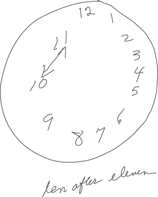
Constructional apraxia is most common after right parietal damage. In addition, as the design complexity increases, the planning abilities of the frontal lobes become more influential. Constructional abilities are typically assessed by asking the patient to draw to command or copy a design (See Fig. 11.3a and b). Asking patients to draw the state and the location of several cities can also identify impairment in spatial relationships, which is often associated with getting lost and difficulty map reading. Regardless of the method of examination, it is best to vary item complexity (e.g., square, diamond inside a square, clock). Clock drawing is an example of a more complex design, and planning problems are evidenced by poor organization of the placement of the numbers around the clock face. Constructional apraxia can be worsened by hemispatial neglect with a tendency to not paying attention to the left side of space (see Fig. 11.4). Clock drawing to command is also sensitive to constructional apraxia, but Fig. 11.5 shows an example of impaired clock drawing that reflects impaired planning rather than constructional apraxia due to visuospatial deficits .


Fig. 11.5
Clock drawing. Reflects impaired executive function including poor planning and writing the time below the drawing (“ten after eleven”) before concretely placing the hands at the 10 and the 11 rather than at the 10 and the 2
Dressing apraxia is defined as difficulty dressing, which is most commonly associated with spatial problems. For example, such patients have difficulty differentiating the top or bottom of a garment and the right or left sleeve as well as putting the correct arm in the correct sleeve. This problem is most commonly associated with right hemisphere damage, especially of the parietal lobe.
Stay updated, free articles. Join our Telegram channel

Full access? Get Clinical Tree


