Disorders of Skeletal Muscle

Chapter 79 Disorders of Skeletal Muscle


Disorders of skeletal muscle encompass a variety of illnesses that cause weakness, pain, and fatigue in any combination. They vary from the protean symptoms of aches, cramps, and pains that often defy any explanation to the muscular dystrophies, which one recognizes instantly on clinical grounds. The disorders with primary involvement of the anterior horn cells (e.g., amyotrophic lateral sclerosis and the spinal muscular atrophies), neuromuscular junction disorders (myasthenia gravis, Lambert-Eaton syndrome, and congenital myasthenia), and certain polyneuropathies (e.g., chronic inflammatory demyelinating polyneuropathy) can cause similar symptoms and may be difficult to differentiate from primary disorders of muscle on clinical grounds. Some definitions are worth reviewing. Myopathy simply refers to an abnormality of the muscle and has no other connotation. Muscular dystrophies are genetic myopathies usually caused by a disturbance of a structural protein or enzyme, resulting in necrosis of muscle fibers and replacement by adipose and connective tissue. Congenital myopathies are a group of illnesses that usually present in young children; many are relatively nonprogressive. However, rare “congenital myopathies” may manifest initially in adults (e.g., central nuclear myopathy, nemaline myopathy) and can be progressive. With the advent of molecular genetics, we recognize that many are allelic to what others have reported as dystrophies. Myositis implies an inflammatory disorder and is usually reserved for disorders in which the muscle histology shows an inflammatory response. The myotonias are diseases in which the occurrence of involuntary persistent muscle activity accompanied by abnormal repetitive electrical discharges distorts the normal contractile process. This occurs after percussion or voluntary contraction. Metabolic myopathies, in this context, refer mainly to abnormalities of muscle biochemistry that impair the resynthesis of adenosine triphosphate (ATP) or cause an abnormal storage of material in the cell. The term endocrine myopathy refers to myopathies associated with disorders of the thyroid and parathyroid glands and to myopathies associated with corticosteroids.


Striated muscle is the tissue that converts chemical energy into mechanical energy. The component processes include (1) excitation and contraction occurring in the muscle membranes, (2) the contractile mechanism itself, (3) various structural supporting elements that allow the muscle to withstand the mechanical stresses, and (4) the energy system that supports the activity and integrity of the other three systems. The logical categorization of myopathies is according to the part of the system involved. Until recently, this was impossible because the molecular basis of muscle activity was unknown. Scientific advances since the 1980s made this classification possible. Abnormalities in the membrane ion channels (channelopathies) involved in muscle excitation cause various forms of myotonia and periodic paralysis (see Chapter 64). The complex of proteins that include dystrophin, the sarcoglycans, and α-laminin constitute a vital structural mechanism linking the contractile proteins with the extracellular supporting structures. Defects in these proteins are the basis of many forms of muscular dystrophy. Although knowledge remains incomplete, it seems reasonable to modify the classic description of the myopathies to incorporate the new information. For this reason, in the sections that follow, disease descriptions are under the heading of their known molecular defect where possible; the classic appellation appears parenthetically. Before describing the illnesses themselves, we first review the techniques used in the clinical evaluation of patients.


Muscle Histology


The technique of muscle biopsy is not difficult. Under local anesthesia, a small incision made over the muscle allows, with careful dissection, removal of a small strip of muscle. Needle biopsies are useful in some situations. Histochemical studies of frozen sections are essential for proper interpretation. A transverse section of normal muscle shows fibers that are roughly of equal size and average approximately 60 mm in transverse diameter (Fig. 79.1). The muscle fibers of infants and young children are proportionately smaller. Each fiber consists of hundreds of myofibrils separated by an intermyofibrillar network containing aqueous sarcoplasm, mitochondria, and the sarcoplasmic reticulum with the associated transverse tubular system. Surrounding each muscle fiber is a thin layer of connective tissue (the endomysium). Strands of connective tissue group fibers into a fascicle, separated from each other by the perimysium. Groups of fascicles are collected into muscle bellies surrounded by epimysium.


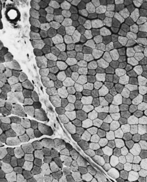
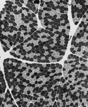
Situated at the periphery of the fibers are the sarcolemmal nuclei. The fibers are of different types. The simplest division is into type 1 and type 2 fibers, best demonstrated with the histochemical reaction for myosin adenosine triphosphatase (ATPase) (Fig. 79.2). The type 1 and type 2 fibers are roughly equivalent to slow and fast fibers or to oxidative and glycolytic fibers in human muscle. The best demonstration of the intermyofibrillar network pattern is with the histochemical reactions for oxidative enzymes, such as reduced nicotinamide adenine dinucleotide dehydrogenase. A regular network extends across the whole fiber. In addition to the routine stains with hematoxylin and eosin, modified Gomori-trichrome, myosin ATPase, and nicotinamide adenine dinucleotide dehydrogenase, the use of other special stains demonstrates fat (Sudan black or oil red 0), complex carbohydrates (periodic acid–Schiff), amyloid (Congo red), or specific enzymes (e.g., phosphorylase, succinic dehydrogenase, cytochrome oxidase). Immunocytochemical techniques demonstrate the location and integrity of structural proteins such as dystrophin. They also characterize cell types in biopsy samples with inflammatory changes.


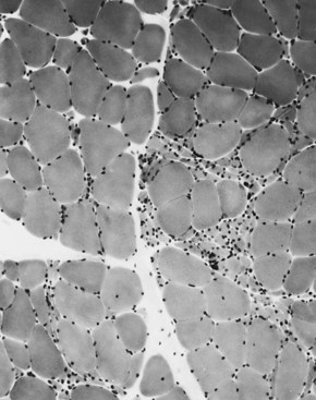
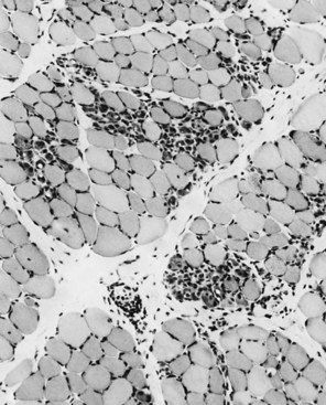
Changes of Denervation


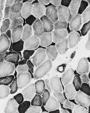
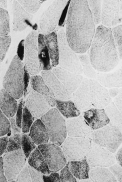
When muscle loses its nerve supply, muscle fibers atrophy, often resulting in fiber squeezing into the spaces between normal fibers and assuming an angulated appearance (Fig. 79.3). Scattered angulated fibers appear early in denervation. Sometimes, picturesque changes in the intermyofibrillar network occur, as in the target fiber, which characterizes denervation and reinnervation. This is a three-zone fiber on which the intermediate zone stains more darkly, and the central “bull’s eye” stains much lighter than normal tissue (Fig. 79.4). Often a neighboring nerve twig reinnervates a denervated fiber. This results in the same anterior horn cell supplying two or more contiguous fibers. If that nerve twig then undergoes degeneration, instead of only one small angulated fiber being produced, a small group of atrophic fibers develops. Group atrophy suggests denervation (Fig. 79.5). As the process continues, large groups of geographical atrophy occur in which entire fascicles are atrophic. In addition to the change in size, a redistribution of the fiber types occurs as well. Normally a random distribution of type 1 and 2 muscle fiber types exists, sometimes incorrectly called a checkerboard or mosaic pattern. The same process of denervation and reinnervation results in larger and larger groups of contiguous fibers supplied by the same nerve. Because all fibers supplied by the same nerve are of the same fiber type, groups of type 1 fibers next to groups of type 2 fibers replace the normal random pattern. This fiber type grouping is pathognomonic of reinnervation (Fig. 79.6). When long-standing denervation is present, the atrophic muscle fibers almost disappear, leaving small clumps of pyknotic nuclei in their place.
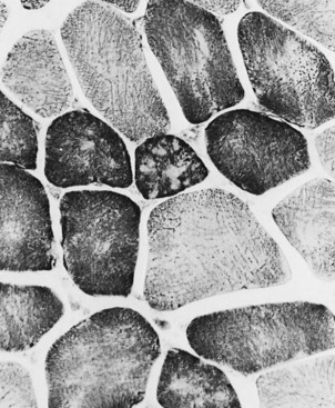


Myopathic Changes


Primary diseases of the muscle cause much greater variation in pathological changes than denervation. The type of change occurring depends on the type of muscle disease. The normal peripherally placed nuclei may migrate toward the center of the fiber. Internalized nuclei may be seen in normal muscle (up to 2% of fibers), but when they are numerous, they usually indicate a myopathic process. Numerous internal nuclei are a feature of the myotonic dystrophies and the limb-girdle muscular dystrophies (LGMDs). Occasionally, one sees internal nuclei in some of the chronic denervating conditions (e.g., juvenile spinal muscular atrophy). Necrosis of muscle fibers, in which the fiber appears liquefied and later presents as a focus of phagocytosis, occurs in many of the myopathies. These changes usually represent an active degenerative process. They often are a feature of myoglobinuria, toxic myopathies, inflammatory myopathies, and metabolic myopathies and are also seen in dystrophies. Fiber-size variation may occur in primary diseases of muscle, with large fibers and small fibers intermingling in a random pattern. It is sometimes the only indication of a pathological process. Fiber splitting often accompanies muscle fiber hypertrophy. In transverse section, recognition of split fibers is by a thin fibrous septum, often associated with a nucleus that crosses partway but not all the way across the fiber. A detailed study of serial transverse section may reveal more split fibers than in a single section. Fiber splitting is particularly visible in dystrophic conditions such as LGMD, but not usually a feature of Duchenne muscular dystrophy (DMD), Becker muscular dystrophy (BMD), or acquired myopathies such as polymyositis.


Degeneration and regeneration of fibers characterize many illnesses. When this occurs, the regenerating fibers often become basophilic, and myonuclei enlarge because of the accumulation of ribonucleic acid (RNA) needed for protein synthesis. Fiber basophilia is a sign of an active myopathy. It is particularly characteristic of DMD, in which small basophilic groups of fibers may be prominent. Cellular responses include frank inflammatory reactions around blood vessels, which characterize the collagen vascular diseases and dermatomyositis. Endomysial inflammation with invasion of non-necrotic muscle fibers occurs in inclusion body myositis and polymyositis. Importantly, pronounced inflammatory cellular responses may occur in dystrophies, particularly facioscapulohumeral muscular dystrophy and dysferlinopathies. Even the so-called congenital inflammatory myopathies actually represent forms of congenital muscular dystrophy.


Fibrosis is another reactive change in muscle. Normally a very thin layer of connective tissue separates the muscle fibers. In dystrophic conditions, this layer thickens, and muscle fibrosis may be quite pronounced. In DMD, muscle fibrosis gives the muscle a hard, gritty texture; this texture also occurs in some congenital dystrophies. In the inflammatory myopathies, there may be a loose edematous separation of fibers, but fibrosis is not usually characteristic of the active phase of the disease except where associated with systemic sclerosis.


Changes in the intermyofibrillar network pattern are common in myopathic disorders. There is often a moth-eaten, whorled change to the intermyofibrillar network in LGMD and facioscapulohumeral dystrophy (FSHD) (Fig. 79.7); the intermyofibrillar network loses its orderly arrangement and swirls, resembling the current in an eddying stream. These changes may be seen in several diseases but tend to be much more common in the myopathies.


Other Changes


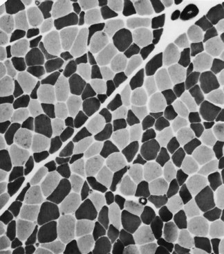
Selective changes in fiber types occur. Type 2 fiber atrophy is one of the most common abnormalities seen in muscle (Fig. 79.8). Type 2 atrophy, particularly if limited to type 2B fibers, is nonspecific and indicates muscle disuse. If a limb is casted and the muscle examined some weeks later, selective atrophy of type 2 fibers is noted. Any chronic systemic illness tends to produce type 2 atrophy. It occurs in rheumatoid arthritis, nonspecific collagen vascular diseases, cancer (hence the name cachectic atrophy), mental retardation in children, and pyramidal tract disease. Type 2B fiber atrophy can also result from chronic corticosteroid administration. Therefore, type 2 fiber atrophy should probably be regarded as a nonspecific result of anything less than robust good health.


Type 1 fiber atrophy is more specific. It occurs in some of the congenital myopathies and dystrophies, congenital myasthenia, and is characteristic of myotonic dystrophy type 1. Changes in the proportion of fibers in the biopsy are quite separate from changes in the fiber size. The name fiber type predominance refers to a change in the relative numbers of a particular fiber type. Type 1 fiber predominance is a normal finding in the gastrocnemius and deltoid muscles. It is also the hallmark of congenital myopathies and many of the early dystrophies. Type 2 fiber predominance is seen in the lateral head of the quadriceps muscle. Type 2 predominance occurs occasionally in juvenile spinal muscular atrophy and motor neuron disease but is not firmly associated with any particular disease condition.


Some changes in muscle biopsy results are pathognomonic of a particular disease. Thus, perifascicular atrophy, in which the atrophic fibers are more numerous around the edge of the muscle fascicles, is the hallmark of dermatomyositis. The presence of lipid vacuoles or abnormal pockets of glycogen characterize the various metabolic myopathies. Enzyme defects including phosphorylase deficiency, phosphofructokinase (PFK) deficiency, and myoadenylate deaminase deficiency are detectable with appropriate histochemical stains. Interpretation of muscle biopsy usually includes the description of a constellation of changes and the subsequent association of these changes with a particular diagnosis. Illnesses that have characteristic biopsies include infantile spinal muscular atrophy, dermatomyositis, inclusion body myositis, the congenital myopathies, lipid storage myopathies, and glycogen storage diseases (e.g., Pompe disease, phosphorylase deficiency). Immunocytochemical staining can also distinguish many forms of muscular dystrophy from each other. Although not disease specific, characteristic biopsy changes differentiate chronic denervation from acute simple denervation.


Immunohistochemistry and Immunoblot


The use of biopsy material to identify missing proteins is increasing with the greater availability of commercial antibodies to the proteins of interest. If deoxyribonucleic acid (DNA) studies are unremarkable in a patient with DMD, the diagnosis rests on demonstrating absent or abnormal dystrophin in the tissue. All the sarcoglycans are demonstrated using similar techniques. Deficiency of any of the sarcoglycans causes a muscular dystrophy, and because they comprise a complex, when one is missing, all or some of the others may be absent in the biopsy. The α-sarcoglycan is particularly prone to be missing, which makes it a suitable and economical screening tool. Absence or reduction of laminin-α2 chain (merosin) or α-dystroglycan occurs in some forms of congenital muscular dystrophy. The lack of nuclear membrane staining with anti-emerin antibodies occurs in X-linked Emery-Dreifuss muscular dystrophy. Histochemical studies are useful to look for several proteins such as desmin, ubiquitin, and amyloid. In addition, biopsy material may give information about the type of inflammatory cell and the affinity for various markers such as CD68 (macrophages and dendritic cells), CD20 (B cells), CD3 (activated T cells), CD8 (cytotoxic T cells), and CD4 (T-helper and dendritic cells); these identify cells involved in cytotoxic, humoral, and innate immune mechanisms. Antibodies to the membrane attack complex may demonstrate the cells marked for destruction by the immune process, as in the vascular endothelium in dermatomyositis.


Immunoblot or Western blot of muscle biopsy is more sensitive than immunohistochemistry, particularly when dealing with an enzyme deficiency or nonstructural protein in evaluation of dystrophies. Patients with Becker muscular dystrophy may have normal-appearing immunostaining for dystrophin, because the commercial antibodies may react to that part of the dystrophin protein that is normally made. However, immunoblot reveals abnormal size or amount of dystrophin in such cases. Immunoblotting is valuable in assessing for calpainopathy (LGMD2A), dysferlinopathy, LGMD2B, and in the secondary α-dystroglycanopathies.


Specific Disorders


Muscular Dystrophies


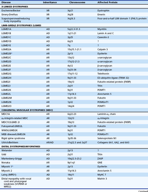
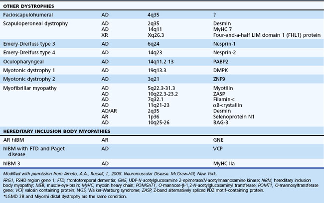
The muscular dystrophies are a group of hereditary muscle disorders that occur at all ages and with varying degrees of severity. The traditional classification is on clinical grounds. Increasing information about the molecular basis of these disorders provides both reassurance and puzzlement to clinicians (Table 79.1). Different dystrophies are due to distinct molecular abnormalities; however, patients with similar molecular defects may show a wide variability in phenotype not always easily explained.


For the most part, the underlying molecular abnormalities in the dystrophies involve structural proteins. Therefore, it is useful to review these proteins as they occur in normal muscle. The contractile proteins, actin and myosin, are arrayed with other proteins such as troponin to form the familiar thick and thin filaments of the sarcomere. The reaction between actin and myosin results in realignment between the two molecules. In the sliding filament model, the thick and thin filaments form an array that slides back and forth.


The contractile proteins connect to the “outside” of the cell by means of a complex of proteins that ultimately links up with the basal lamina. The first step in this connection is the protein, dystrophin, located on the cytoplasmic face of the muscle membrane. This large protein (427 kD) is coded by a gene on the short arm of the X chromosome. Dystrophin relates to spectrin and other structural proteins and consists of two ends separated by a long, flexible rodlike region. The amino terminus binds to the actin molecule, and the carboxyl terminus, which is rich in cysteine, links dystrophin to a complex of glycoproteins in the sarcolemma. Two of these, the dystroglycans, form a direct link between dystrophin and part of the laminin molecule. α-Dystroglycan is a 156-kD protein located outside the membrane and linked to the laminin-α2 chain. It also connects with β-dystroglycan, which is a 43-kD transmembrane component of the complex and links with dystrophin. The other glycoproteins are the sarcoglycans, of which there are four known at present, labeled alphabetically α- (50 kD), β- (43 kD), γ- (35 kD), and δ- (35 kD) sarcoglycans. All span the sarcolemmal membrane, but their relationship to the dystroglycans and their precise function are unclear. Coding of the sarcoglycans is on different autosomal chromosomes. The α2 chain of laminin (merosin) provides the anchor into the extracellular matrix because it is via the globular domain of this part of the molecule that α-dystroglycan attaches to laminin. Merosin also binds to a7β1D integrin, a protein complex located on the sarcolemma membrane. Dystrophin, the sarcoglycans, the dystroglycans, and merosin appear to function as a unit in stabilizing the muscle membrane. Together these proteins make up the dystrophin-glycoprotein complex.


Other sarcolemmal proteins not directly linked to the dystrophin-glycoprotein are also affected in certain forms of muscular dystrophies (e.g., dysferlin, caveolin-3). Also, sarcomeric proteins (e.g., myosin, actin, tropomyosin, myotilin, ZASP [Z-band alternatively spliced PDZ motif-containing protein], filamin-c, desmin, titin, and telethonin, etc.), important in stabilizing the contractile apparatus, are mutated in certain types of dystrophies. Mutations of the muscle-specific calcium-dependent protease, calpain-3 gene, are responsible for the majority of non-dystrophin-related LGMDs in patients of Italian and Spanish ancestry. In addition, secretory enzymes (e.g., O-mannose-β-1,2-N-acetylglucosaminyl transferase, fukutin, and fukutin-related protein), which probably play a role in glucosylation of α-dystroglycan and other import proteins, are responsible for some forms of congenital muscular dystrophy. Mutations in fukutin-related protein also cause LGMD2I, the most common form of non–dystrophin-related dystrophy in patients from northern Europe. Furthermore, mutations encoding for the nuclear envelope proteins, emerin, lamin A/C, and nesprin 1 and 2 cause Emery-Dreifuss muscular dystrophy.


Dystrophin Deficiency (Duchenne Muscular Dystrophy, Becker Muscular Dystrophy, and Atypical Forms)


An absence or deficiency of dystrophin is responsible for two disorders that cause progressive destruction of muscle. The responsible gene is located on the short arm of the X chromosome at locus Xp21. The gene is extremely large, comprising more than 2.5 million base pairs and 79 exons or coding regions. Approximately two-thirds of cases are associated with a detectable deletion or duplication of segments within the gene. The others are presumably due to point mutations too small to be detected using standard techniques. “Hot spots” for these gene deletions exist, notably between exons 43 and 52 and particularly 44 and 49 (Nobile et al., 1997). Whether the deletion is in frame or out of frame (see Chapter 40) determines whether dystrophin is absent from the muscle or present in a reduced altered form. This has clinical significance because the former is usually associated with the severe Duchenne variety of the disease (DMD), whereas the latter may cause the milder Becker variant (BMD). In BMD, the abnormal dystrophin preserves enough function to slow down the progress of the illness. Reading of the DNA code is triplet by triplet. Maintenance of this reading frame throughout the length of the gene is required for dystrophin production. If a deletion removes a multiple of three base pairs, the reading frame may be intact upstream and downstream and may make limited sense, as if the sentence “You cannot eat the cat” were changed to “You not eat the cat,” and some modified dystrophin may be formed. This is often the situation in the mild form of dystrophin deficiency. In the severe form, the reading frame is destroyed, as if a deletion resulted in the sentence “Yoc ann ote att hec at.” Exceptions to this rule exist, and frameshift deletions have been associated with the milder form of the disease, particularly at the 5′ end of the gene in exons 3 to 7.


The prevalence of DMD in the general population is approximately 3 per 100,000, and the incidence among liveborn males is 1 per 3500. BMD is approximately one-tenth as common. Although the inheritance is clearly X-linked recessive, almost a third of cases are sporadic. Presumably this is due to a spontaneous mutation occurring either in the child or in the mother’s ova.


It is not difficult to imagine that an absence of dystrophin would severely impair the integrity of the sarcolemmal membrane. Attention previously focused on this membrane because of electron microscopic (EM) evidence that it contains breaches associated with wedge-shaped areas of destruction in the adjacent muscle cell. One assumes that the absence of a supporting protein renders the membrane susceptible to mechanical damage. This means that molecules such as calcium would have unlimited access to the fiber and initiate a whole chain of destructive processes, producing necrosis of the muscle fiber. The process would then involve continual degeneration, with repeated attempts at regeneration on the part of the surviving satellite cells. Eventually, however, this process leads to severe loss of muscle and replacement of the muscle fibers with fibrous tissue.


Several animals have dystrophin deficiency; the better known include the mdx mouse and the dog model. The situation in the mouse model is unusual. The animals appear to be relatively normal except for a phase early in life during which pathological abnormalities in the muscle are noted. In the dogs, however, the dystrophin deficiency is associated with obvious weakness, and microscopy reveals abnormal muscle, thus making this animal suitable for the evaluation of potential treatment.


Duchenne Muscular Dystrophy


Even in the severe variety of DMD, affected children are normal at birth. During the second year when the boys begin walking, the clumsiness seen in all toddlers persists. Soon the children have to place one hand on the knee to assume an upright position when rising from the floor (Gower maneuver). Often at this stage, the calf muscles are rather firm and rubbery (pseudohypertrophy) (Fig. 79.9). Within 2 to 3 years, parents notice that the child runs properly and is never able to jump clear of the floor with both feet. In the absence of therapy, tightness across several joints in the legs is noted. The iliotibial bands and the heel cords are usually the first to become tight. This is particularly noticeable in boys who habitually walk on their toes.
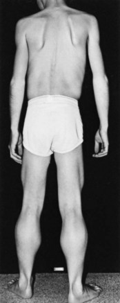


By 5 or 6 years of age, labored stair climbing is the rule, and moving upward requires use of the railing. Sometime between the ages of 2 and 6, there will be a period of apparent improvement when the child gains motor skills. This is illusory because it simply represents the child’s natural development, which muscle weakness has not yet outpaced. At the age of 6 or 7, the boys often complain of sudden spontaneous falls. At first, these falls occur when the child is in a hurry or knocked off balance by playmates. The fall is quite spectacular to the onlooker; the knees collapse abruptly, and the child drops like a stone to the ground. At approximately 8 to 10 years of age, affected children cease to be able to climb stairs or stand up from the floor, and it is at approximately this time when they begin using a wheelchair for locomotion. Earlier studies suggested that these children began using a wheelchair and lost the ability to walk at about age 9, but in a population treated with bracing, reconstructive surgery, and physiotherapy, the average age of confinement to a wheelchair was 12.2 years. The true natural history of the disease is difficult to ascertain because many physicians and parents put considerable effort into keeping the children straight and the limbs supple.


Contractures of the hips, knees, and ankles become severe when the relatively untreated child spends much of the day in a wheelchair. The hips and knees lock at 90 degrees, and the feet turn downward and inward in an exaggerated position of equinovarus. It is very difficult to get normal shoes to fit them, and it is impossible for them to sleep except in one position: usually with the knees propped up with pillows and slightly turned on one side. Handling the children at this stage becomes very difficult, and back pain and limb pain almost inevitably accompany this severe stage of muscular dystrophy. Development of a severe scoliosis compromises respiratory function.


Characteristic cardiac involvement is degeneration and fibrosis of the posterolateral wall of the left ventricle. Besides the abnormal electrocardiogram (ECG), valve motion, wall thickness, and wall motion are also abnormal. Affected children die from either respiratory failure or a cardiomyopathy that is relatively resistant to treatment.


The least invasive test to confirm the diagnosis is to obtain DNA studies looking for a deletion in the dystrophin gene. In the 30% of patients without a deletion, a muscle biopsy is necessary to establish the absence of dystrophin. Three antibodies are available against the ends (Dys-2 for the carboxyl terminus and Dys-3 for the amino terminus) and the rod region (Dys-1) of the molecule. Absence of the amino terminus, the end that binds with actin, appears to be associated with the more severe symptoms. In dystrophin deficiency, the protein is absent or the stain is irregular and fragmented. Approximately 1% of fibers demonstrate a rim of dystrophin. These are the revertant fibers, in which the gene has undergone another mutation, putting the coding sequence back in-frame. Where doubt exists, immunoblotting shows a decreased amount of dystrophin in the tissue.


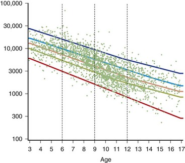
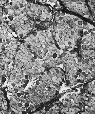
The serum concentration of creatine kinase (CK) is markedly elevated in this illness; levels greater than 10,000 mU/mL are common. Electromyography (EMG) shows myopathic changes (see Chapter 32B), and muscle biopsy demonstrates variation in the size of fibers, fibrosis, groups of basophilic fibers, and opaque or hypercontracted fibers (hyaline fibers) (Figs. 79.10 to 79.12).


Treatment of Duchenne Muscular Dystrophy


Bracing

The appropriate use of bracing may delay the child’s progression to a wheelchair by approximately 2 years. A major factor responsible for inability to stand or walk is weakness of the quadriceps. Such weakness causes the knee to collapse when even slightly flexed; the only stable position is in hyperextension. The boy is then reluctant to bend the knee in walking and may remain rooted to the ground, unable to move the feet. The addition of a long-leg brace (knee-foot orthosis) can help solve this problem. Such a device stabilizes the knee and prevents the knee from flexing. The children walk stiff legged but do not have the same problem with falling they had previously. Generally, children are ready for bracing when they have ceased to climb stairs, are having great difficulty arising from the floor, and are having frequent daily falls. On examination, a knee extensor muscle that is unable to straighten the knee against gravity is also an indication for bracing. One often hears the comment that the weight of the brace makes it difficult for the child to walk. Because the brace functions as a pendulum, and slight elevation of the hip is sufficient to bring the leg forward, the weight of the brace is rarely a problem. There may be some advantage to a lightweight plastic knee-foot orthosis, but difficulty exists to keep the foot straight with such a device, whereas the high-top boot worn with the double-upright brace provides excellent stability. The choice between plastic and metal often comes down to personal preference of the patient or physician.


Surgery

Reconstructive surgery of the leg often accompanies bracing. Indeed, for most children, the two occur during the same hospital admission. The purpose of leg surgery is to keep the leg extended and prevent contractures of the iliotibial bands and hip flexors. Shortening of the iliotibial bands is associated with a stance in which the boy’s legs are widely abducted. As noted before, the long-leg brace acts like a pendulum. If the foot is widely abducted, the child cannot swing the leg forward. The only effect of lifting the hip in this case is that the leg tries to swing inward toward the midline. This is impossible because the abduction is due in the first place to the resistance of the iliotibial band contractures. A simple way to maintain function in the leg is to perform percutaneous tenotomies of the Achilles tendons, knee flexors, hip flexors, and iliotibial bands. This procedure often allows a child who is becoming increasingly dependent on a wheelchair to resume walking.


Modern techniques of spinal stabilization are an important technique for DMD patients. Because of the extreme discomfort of a severe scoliosis and the respiratory problems associated with it, spinal surgery is an acceptable procedure for patients in managing the late stages of the disease. Consider surgery in patients with 35 degrees or more of scoliosis and significant discomfort. To reduce the risks associated with surgery, forced vital capacity ideally should be greater than 35% of predicted.


Gene Therapy

In theory, the treatment and cure of muscular dystrophies is replacement of the defective gene. The disease is due to a negative effect, the lack of dystrophin, not to a positive effect due to the presence of a toxic gene product. The possibility of replacing old muscle with new is enticing and has occupied researchers for the past decade. The first attempt was to use normal myoblasts grown from unaffected muscle. Myoblasts injected into muscle were to fuse with the dystrophic muscle and carry the normal gene with them. Despite dystrophin expression in some of the muscle fibers, the low percentage of fibers fused provided no clinical effect. The reasons are many: injected cells diffuse only a short distance, they are prone to rejection, and the surviving muscle proves resistant to fusion with these myoblasts. Attempts are ongoing to circumvent these drawbacks, but so far, myoblast transfer is unproven.


More promising is the attempt to insert the dystrophin gene into a vector that can carry the gene into the muscle. Several problems exist. The large size of the gene makes it difficult to insert in the usual vectors. An abbreviated version of the full gene may be satisfactory, but mildly affected patients exist in whom such truncated genes occur naturally. The gene will have to work in conjunction with a promoter that allows dystrophin production to be limited to the muscle. The viral vector must also be safe. This requires removal of some or all of the viral genes. At present, the adenovirus is the front runner from which a vector might be developed, but other viruses (e.g., retroviruses) are a consideration. Human studies are in a planning stage.


Another possible treatment is the use of drugs that allow for “read-through” of stop codons such as gentamicin and PTC-124. However, clinical trials of these agents have demonstrated no significant clinical benefit. Studies are underway to assess the utility of antisense oligonucleotides that have the ability to bring the reading frame back “in frame.”


Becker Muscular Dystrophy


BMD shares all the clinical characteristics of the severe form of DMD but has a milder course. The disease begins in the first decade, although parents often notice the first signs of weakness later because of the milder symptoms. Occasionally, symptom onset is delayed until the fourth decade or later. The muscular hypertrophy, contractures, and pattern of weakness are similar to that seen in DMD. These boys, however, continue to walk independently past the age of 15 years and may not use a wheelchair until they are in their 20s or even later. Frequent complaints in teenagers with BMD are leg cramps and other muscle pains, which are often associated with exercise and are more severe than in DMD. A significant proportion of these patients have a cardiomyopathy that can be more disabling than the weakness. Cardiac transplantation has been very successful in some patients with this form of the illness.


As with DMD, serum CK levels are elevated but not typically as high. EMG demonstrates myopathic features. These abnormalities are nonspecific, however, and diagnosis requires demonstration of a mutation in the dystrophin gene or reduced quantity or size of dystrophin on muscle biopsy. Immunostaining of muscle tissue with Dys 1, Dys 2, and Dys 3 antibodies may or may not demonstrate an abnormal staining pattern. Therefore, definitive diagnosis often requires quantitation of the dystrophin in the muscle with immunoblot (Western blot) studies.


Because boys do not have much trouble in the first few years, there is a reduced need for aggressive physiotherapy, surgical reconstruction, and night splints. Patients with BMD are less prone to develop kyphoscoliosis, perhaps because they lack wheelchair confinement until after the spine has become fully mature. We have used corticosteroids only occasionally in patients with BMD. The stabilizing effect of steroids is less noticeable when the disease is already fairly stable. In every other respect, including bracing and genetic counseling, disease treatment is the same as that for the severe form.


Genetic Counseling


Because DMD is an X-linked recessive disorder, we recommend ascertainment of the carrier state of all women related to an affected person by maternal linkage. Up to 30% of cases may be sporadic and due to new mutations or deletions. In the experience of many clinicians, an even higher percentage of new patients arriving in the clinic are sporadic cases, perhaps because genetic counseling is widely available and the women who carry the abnormal gene decide not to have children.


Most female carriers are asymptomatic, but approximately 8% manifest weakness and have a clinical phenotype similar to BMD. Manifesting carriers will usually have an elevated serum CK level and myopathic EMG. Muscle biopsy will usually demonstrate a mosaic pattern or patchy staining of dystrophin on the sarcolemma. However, these laboratory tests and muscle biopsy are insensitive in diagnosing carrier status in asymptomatic females, and diagnosis of carrier status requires genetic studies.


Genetic analysis of all potential carriers is advisable. In a family in which the disease is associated with a deletion, there is little problem in determining whether the woman is carrying the affected X chromosome, using techniques that are presently available. Genetics laboratories now have the ability to identify the presence of a mutant gene over the background contributed by the normal allele. This involves an analysis of the gene “dosage,” comparing two normal alleles that have a double dose against a deleted allele and a normal allele that have a single dose (Voskova-Goldman et al., 1997). The basis for current diagnosis of carrier status after identification of a deletion in a proband is by analysis of a gene dosage. Our diagnostic strategy uses fluorescence in situ hybridization (FISH) to detect female carriers with major deletions in the dystrophin gene. Unfortunately, situations exist in which a mutation is identified in a boy with “sporadic” DMD but not in the mother, and yet the mother is still a carrier. This occurrence is secondary to germline mosaicism in which the mutation in the mother lies only in a percentage of her oocytes. The estimated recurrence rate of DMD is as high as 14% even in such cases. Prenatal diagnosis using amniotic cells or chorionic villus biopsies can identify the affected fetus and, more importantly, those who are unaffected.


Other Limb-Girdle Dystrophies


The most important recent development in muscle disease relates to a group of disorders that were clearly dystrophic but defied proper classification. The traditional diagnosis of LGMDs cloaked the clinician’s uncertainty. In some patients, weakness was generally proximal, but other characteristics were disparate. Some cases were dominantly inherited, others recessively. Some had more hip than shoulder weakness, others the reverse. The illness could be mild in late life, others severe and early. There was general recognition that the rubric LGMD encompassed a group of different illnesses. Beginning with the discovery that a defect in one of the sarcoglycans caused a severe form of dystrophy occurring in North Africa, the delineation of several other entities characterized by defects in structural proteins or enzymes followed. These include the sarcoglycans, the α2 chain of laminin (merosin), calcium-activated protease, calpain-3, and others. The designation of autosomal dominant LGMD is type 1, and the designation of autosomal recessive LGMD is type 2. Subclassification with an alphabetical letter characterizes distinct genetic forms of LGMD1 and LGMD2 (see Table 79.1). The following sections outline the known protein abnormalities and then comment on the more amorphous forms of LGMD. The prevalence of these diseases as a group probably approaches 1 per 100,000 (van der Kooi et al., 1996). The autosomal recessive LGMDs are more common than the autosomal dominant LGMDs.


Autosomal Dominant Limb-Girdle Muscular Dystrophies


LGMD1B (Lamin A/C Deficiency)

This myopathy is allelic with the disorder previously reported as autosomal dominant Emery-Dreifuss muscular dystrophy (EDMD) (Bonne et al., 2000; van der Kooi et al., 1996). Some patients manifest with a limb-girdle pattern of weakness, while others present with a humeral-peroneal weakness and early contractions. Cardiomyopathy with severe conduction defects and arrhythmias may also occur, with or without skeletal muscle involvement. Sudden death secondary to fatal arrhythmias is common, and pacemaker insertion is often necessary. Serum CK levels may be normal or elevated up to 25-fold. Muscle biopsies demonstrate dystrophic features with the rare occurrence of rimmed vacuoles.


LGMD1B is localized to mutations in the lamin A/C gene located on chromosome 1q11-21 (Bonne et al., 1999; van der Kooi et al., 1997). Alternative splicing of the lamin A/C messenger (m)RNA transcript produces lamins A and C. Lamin A/C is an intermediate-size filament located on the nucleoplasmic surface of the inner nuclear membrane, where it interacts with various lamin-associated proteins including emerin, the abnormal protein associated with X-linked EDMD. Lamin A/C may also bind to heterochromatin. Immunostaining of the nuclear membrane with anti-emerin antibodies is normal, helping distinguish this myopathy from X-linked EDMD. Electron microscopy reveals alterations in myonuclei, including the loss of peripheral heterochromatin, altered interchromatin texture, and fewer than normal nuclear pores (Sabatelli et al., 2001).

< div class='tao-gold-member'>

Only gold members can continue reading. Log In or Register to continue

Stay updated, free articles. Join our Telegram channel

Jun 19, 2016 | Posted by in NEUROLOGY | Comments Off on Disorders of Skeletal Muscle

Full access? Get Clinical Tree

Get Clinical Tree app for offline access