Brainstem location
Signs/symptoms
Possible structures
Midbrain
Upward eye movement, superior oblique weakness, and pupil constriction dysfunction
CN III, IV
Contralateral arm and leg weakness
Midbrain or pontine corticospinal tract
Contralateral arm and leg loss of pain and temperature sensations
Spinothalamic pathways
Contralateral arm and leg loss of vibration and proprioception
Medical lemniscus
Coma
Reticular activating center
Pons
Ipsilateral facial numbness, chewing problems
CN V
Diplopia from ipsilateral loss of horizontal eye movement
CN VI
Weak ipsilateral facial muscles
CN VII
Ipsilateral hearing loss, dysequilibrium, vertigo
CN VIII
Disconjugate lateral gaze or skew deviation from internuclear ophthalmoplegia
Medial longitudinal fasciculus
Dysarthria and dysphagia
Pontine corticobulbar tracts
Ataxia
Pontine corticopontocerebellar tracts
“Locked-in syndrome”
Central pontine myelinolysis
Medulla
Weak pharyngeal muscles
CN IX
Parasympathetic dysfunction
CN X
Laryngeal muscle weakness
CN XI
Poor and weak tongue movement
CN XII
Ipsilateral Horner’s syndrome
Medullary sympathetic pathway
Vertigo
Vestibular nucleus
Quadriplegia
Bilateral corticospinal tracts at decussation
Blood supply to the brainstem and cerebellum comes from both vertebral arteries and the basilar artery (Fig. 8.1). There are many small penetrating arterioles that enter the brainstem from these major vessels. The arterioles generally supply one side of the medial brainstem (paramedian arteriole) or one lateral side (circumferential arteriole). Three arteries (superior cerebellar artery, anterior inferior cerebellar artery, and posterior inferior cerebellar artery) supply the cerebellum with blood and also may have branches to the brainstem.
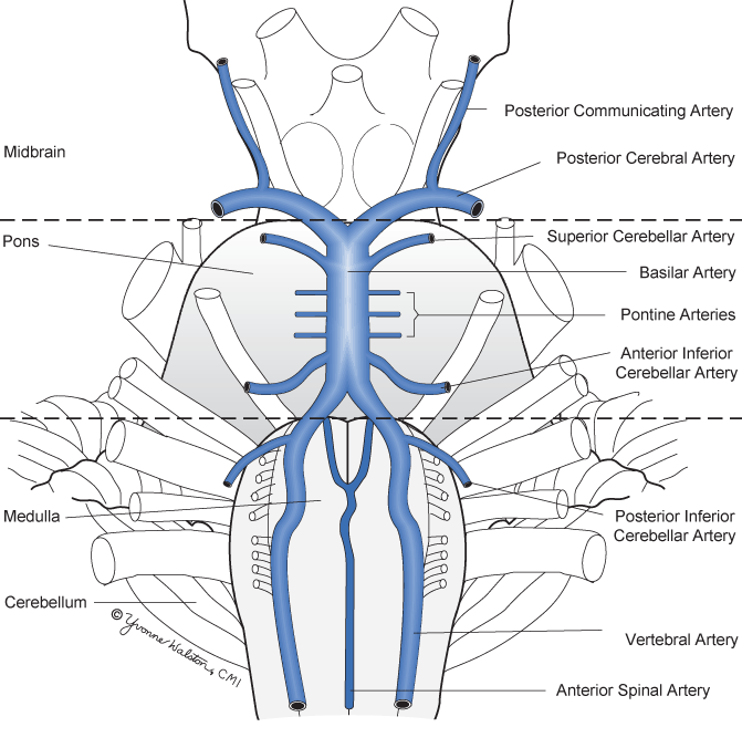
Fig. 8.1
Brainstem and cerebellar circulation
A variety of diseases affect the brainstem but with a lower frequency than the same diseases affecting other brain regions. Brainstem tumors are most often astrocytic and slower growing than astrocytic tumors in the cortex. Bacterial abscesses are rare and most viruses causing encephalitis involve the brainstem less intensely. Hemorrhages involving the brainstem are uncommon. Ischemic strokes of the brainstem occur as lacunes or occlusions of penetrating brainstem arteries and are the most common brainstem disease. However, brainstem strokes are less common than hemispheric or basal ganglia strokes.
The cerebellum occupies about 10 % of the brain volume but contains more neurons than the entire rest of the brain. The cerebellum is divided into the three functional divisions of spinocerebellum, cerebrocerebellum, and flocculonodular lobe (Fig. 8.2). Each division in the cerebellar cortex sends Purkinje cell axons to specific deep cerebellar nuclei and has different functions (Table 8.2). Most input to the highly organized and redundant cerebellar cortex comes from many brainstem nuclei via excitatory mossy fibers that terminate on myriads of granule cells. These granule cell neurons then send inhibitory impulses to Purkinje cells. The inferior olive also sends excitatory input directly to Purkinje cells. Purkinje cells, the only output of the cerebellar cortex, send inhibitory impulses via a GABA neurotransmitter to neurons in the deep cerebellar nuclei that then send output to the brainstem and cerebral cortex.
Table 8.2
Cerebellar functions by location
Function | Cerebellar origin | Deep cerebellar nucleus | Cerebellar efferent connections |
|---|---|---|---|
Adjust the ongoing movements of axial and proximal limb muscles | Spinocerebellum | Fastigial nucleus | Brainstem reticular formation, vestibular nuclei, and motor cortex |
Adjust the ongoing movements of distal limb muscles | Intermediate spinocerebellum | Interposed deep nuclei | Red nucleus and motor cortex |
Initiation, planning, and timing of motor movements | Cerebrocerebellum | Dentate nucleus | Red nucleus and premotor cortex |
Axial control and vestibular reflexes | Flocculonodular lobe | Direct connection to brainstem | Brainstem vestibular nuclei |
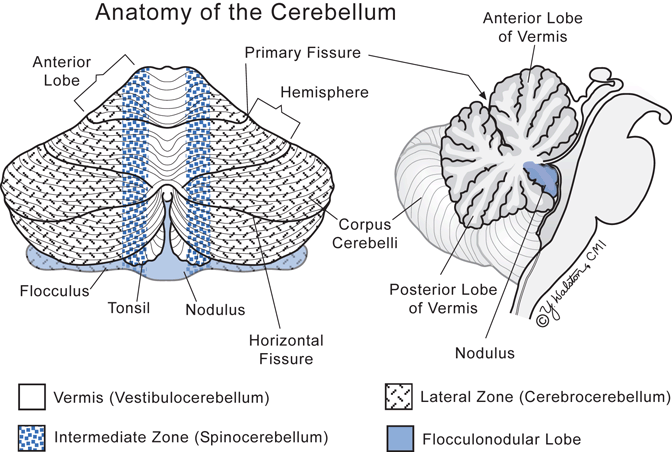
Fig. 8.2
Cerebellar divisions
Cerebellar neurons do not directly produce motor movements but act more as a comparator that compensates for errors in movement by comparing intention with performance and making subtle adjustments. As such, patients with cerebellar diseases do not have weakness or sensory loss. Cerebellar clinical problems are expressed as impaired coordination, imbalance, and even vertigo (Table 8.3). Dysfunction of midline cerebellar structures (vermis of spinocerebellum) produces imbalance problems of midline body structures such as gait and truncal ataxia while cerebellar hemisphere dysfunction produces ataxia of limbs. Unlike the cerebral cortex, damage to one cerebellar hemisphere produces ipsilateral dysfunction.
Table 8.3
Signs suggestive of cerebellar dysfunction and likely cerebellar localization
Vermis (midline cerebellum) |
|---|
Gait ataxia |
Clumsy, uncertain, irregular, staggering steps in walking with wide-based stance like “being drunk.” Tendency to fall to involved side |
Truncal ataxia |
Inability to balance in sitting position at edge of table |
Saccadic eye movement abnormalities |
Conjugate eye movement rapidly to a target results in overshooting of eyes followed by over corrections until target is reached |
Cerebellar hemisphere |
Signs are ipsilateral to side of cerebellar lesion and more abnormal with fast limb movements than slow movements |
Hypotonia |
Diminished arm and leg muscle tone give a loose feeling when passively moving the limb |
Dysdiadochokinesis |
Irregular, uncoordinated rapid movements of hands or fingers. Often tested by asking patient to pat one palm alternately with the palm and dorsum of the opposite hand as rapidly as possible |
Dysmetria |
Inaccuracies in judging distance and target when moving limb to a target with eyes closed |
Cerebellar tremor |
Intention tremor develops when moving arm or leg that is perpendicular to the direction of the movement and often amplifies as the target is reached. Usually tested by asking patient to touch a target and then move quickly to his nose or to lift one heel and place it on the opposite knee and then move heel down the shin |
Ataxic dysarthria |
Poor coordination of articulation resulting in slow, explosive speech |
Flocculonodular lobe |
Nystagmus
Related posts:Stay updated, free articles. Join our Telegram channel
Full access? Get Clinical Tree
 Get Clinical Tree app for offline access

|


