Fig. 12.1
Illustration of surround inhibition. The brain produces the desired motor pattern while simultaneously creating a surround inhibition of competing motor movements
Movement disorders or extrapyramidal disorders are diseases characterized by excessive or abnormal movements in conscious patients. Damage to or presumed dysfunction of the basal ganglia and their brainstem and cerebellar connections is implicated in the etiology of these diseases. The abnormal movements may be the only manifestation of a disease process or part of a constellation of deficits in others. Movement disorders are characterized by either excessive (hyperkinetic) or reduced (hypokinetic) activity. Parkinson’s disease is the classic hypokinetic movement disorder with reduced voluntary and involuntary movement. Chorea, tremor, myoclonus, and tics represent hyperkinetic movement disorders, which cause extra involuntary movement and sometimes interfere with normal voluntary movement as well.
In this chapter, we will first cover definitions of hyperkinetic movement disorders, followed by examples of common movement disorders (Essential tremor, Parkinson’s disease and Huntington disease) .
Hyperkinetic Movement Disorders
When observing a patient with a hyperkinetic disorder, a neurologist notes the topography, symmetry or asymmetry, velocity, task-specificity or posture-specificity of the movement. In addition, the neurologist questions the patient regarding how the movement interferes with voluntary movement, and whether the movement is suppressible, precipitated by any factors, and/or ameliorated by any interventions.
Chorea
Irregular, unpredictable, brief, involuntary jerking movements involving shifting muscles or muscle groups involving the arms, hands, legs, tongue or trunk .
Dystonia
Sustained muscle contractions causing twisting, repetitive movements or abnormal posture . In adults, dystonia is focal in presentation, affecting the neck, the eyes, the jaw or a limb. In children, dystonia can present focally and then generalize over time. The contractions can be painful and may be disabling.
Athetosis
Distal, slow, writhing form of dystonia. It can be seen in combination with chorea .
Ballismus
Uncontrollable, proximal, flinging movements of a limb that is often due to a lesion in the subthalamic nucleus .
Tics
Abrupt, brief, repetitive, stereotypical movements of face, tongue and limbs or vocalizations that may be briefly voluntarily suppressed but is often then followed by a burst of tics when the suppression is removed.
Myoclonus
Sudden, shock-like movements or a pause in movement. The movements can be restricted to a specific muscle or group of muscles or may be multifocal occurring at the same or different times. Myoclonus can be generated in the motor cortex, subcortical areas, brainstem (reticular myoclonus), or spinal cord (propriospinal myoclonus). Many healthy individuals experience myoclonus on falling asleep in the form of a hypnic jerk.
Tremor
A tremor is an involuntary oscillation of a body part caused by alternating contractions of reciprocally innervated muscles. Physiologic tremor in present in the limbs but usually is not bothersome. Stimulants or anxiety can enhance a physiologic tremor causing it to be visible and sometimes intrusive. Current evidence suggests that tremors come from alterations in a complex central oscillatory cycle that involves neurons in the basal ganglia, brainstem, and sometimes the cerebellum. Tremors are classified by frequency, their relationship to movement, and location (Table 12.1).
Table 12.1
Tremor type by clinical presentation
Tremor type (other common names) | Characteristics (examples) |
|---|---|
Rest tremor (pill-rolling tremor) | Present at rest with limb supported against gravity |
• Low-frequency (3–5 Hz), medium to high amplitude (parkinsonian tremor) | |
Action tremor (postural tremor) | Present when voluntarily maintaining a limb still against gravity such as holding arms outstretched |
Usually bilateral but may be asymmetric | |
Low to medium amplitude and medium to high frequency (4–8 Hz) | |
Present during voluntary movements such as writing or eating (essential tremor, enhanced physiologic tremor) | |
Intention tremor (cerebellar tremor) | Present during voluntary movement and often perpendicular to direction of movement |
Medium amplitude and low frequency | |
Often amplifies when limb approaches the target or near the face | |
Interferes with coordination (cerebellar-type tremor as seen in finger-to-nose movements) |
Essential Tremor
Introduction
Essential tremor is the most common movement disorder with an estimated 10 million Americans affected. Essential tremor can begin at any age but is more prevalent with increasing age. It is estimated that 4–5 % of people from age 40 to 60 years have essential tremor. Over the age of 60 years, the incidence rate increases and is estimated at 6–9 %. Although activities of daily living such as feeding, drinking, and writing may be difficult, only a small percentage of patients with essential tremor seek medical attention. Essential tremor can be misdiagnosed as Parkinson disease—likewise in a young patient (under 40 years), other rare causes of tremor should be excluded such as Wilson’s disease and medical conditions such as hyperthyroidism.
Pathophysiology
In the majority of patients with essential tremor , there is a positive family history. Genetic studies have identified several genes suggesting that multiple etiologies may account for essential tremor. At present, the actual pathophysiology of how sporadic or genetic cases develop the tremor is unknown, as structural lesions have not been recognized. There is evidence that the generators of essential tremor are widespread in the brain—including the motor cortex, thalamus, cerebellum, and brainstem, such as the inferior olivary nucleus. These are the same centers that control voluntary movement through a thalamocortical relay, and it is hypothesized that the bidirectional nature of the thalamocortical loops could provide the substrate for oscillatory activity to become established—leading to tremor instead of normal voluntary movement.
Major Clinical Features
The characteristic history is one of slowly progressive bilateral tremors of the hands that began in middle age. The tremor is of medium to high frequency, of low amplitude, sustained and is present immediately with arms outstretched (postural tremor), and absent at rest. The tremor severity can range from socially embarrassing to interfering with activities of daily living such as writing and drinking. Occasionally, the tremor also may involve the head, legs, or voice. Patients relate that the tremor worsens with anxiety, coffee, and some medications. About half of patients endorse a history of their tremor transiently improving after drinking alcohol. Patients with essential tremor could have mild gait imbalance and subtle cognitive changes, but in general, the tremor is the predominant symptom. Weakness, sensory loss, or changes in deep tendon reflexes do not occur in essential tremor . Patients should not have features of Parkinson disease. The clinical diagnosis is made based on the history and exam.
Several drugs that may worsen essential tremor or exaggerate a physiologic tremor include stimulants, lithium, levothyroxine, beta-adrenergic bronchodilators, valproate, prednisone, caffeine, and selective serotonin-reuptake inhibitors (SSRI).
Major Laboratory Findings
No laboratory test is diagnostic. Routine blood tests are normal, and neuroimaging of the spinal cord and brain is normal.
Principles of Management and Prognosis
Patients with mild symptoms usually do not require treatment once they are reassured that they do not have Parkinson disease and that the tremor rarely becomes incapacitating. Many patients find that a small amount of alcohol (glass of wine or beer) suppresses the tremor for hours and is useful when entertaining friends. For patients with severe essential tremor or whose occupation is impaired by the tremor, propanolol and primidone have been successful in reducing the tremor severity. Other agents with evidence of efficacy in treating essential tremor include topiramate and gabapentin. In severe tremor, surgical implantation of electrical stimulators in the thalamus or stereotactic thalamotomy may be indicated.
Parkinson’s Disease
Introduction
Parkinson’s disease (PD) affects more than one million Americans, and each year 50,000–60,000 people are diagnosed with the disease. The direct annual cost in the USA is over $ 10 million. Both sexes are equally involved and the incidence climbs exponentially with increasing age to 7 % above age 70 years. Idiopathic PD usually begins above age 50 while patients with young-onset PD can have onset of symptoms as early as age 21–40 years. A subset of those patients with young-onset PD may have a genetic form of the disease. PD has a dramatic impact on quality of life and a marked reduction in life expectancy.
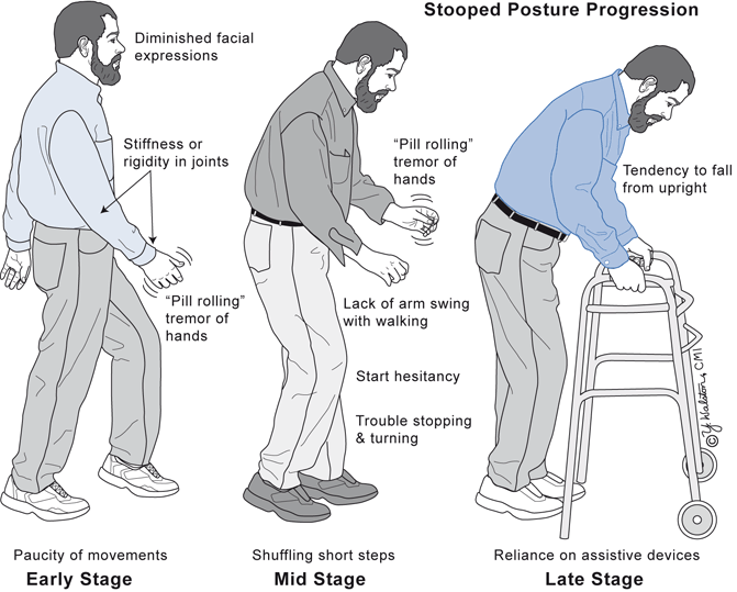
The cardinal symptoms of PD are designated by the abbreviation TRAP: resting Tremor, cogwheel Rigidity, Akinesia or bradykinesia (slowed and small amplitude movements), and Postural instability with gait changes. PD often causes a stooped posture and shuffling gait that are readily apparent even at a distance (Fig. 12.2). PD refers to the primary idiopathic form and represents two thirds of all Parkinsonism. Parkinsonism is the secondary form and refers to the above clinical and biochemical features that develop from specific causes such as repeated head trauma (boxing), infections of the upper midbrain, medications that affect dopamine transmission, or CNS diseases that damage the nigrostriatal pathway and other brain areas .


Fig. 12.2
Postural and gait changes in the progression of Parkinson’s disease
Pathophysiology
Idiopathic PD results from the slowly progressive death of CNS dopaminergic neurons, although what triggers the demise of these specific neurons is not known. Current theories include exposure to environment neurotoxins, abnormal mitochondrial function, abnormal oxidative metabolism, and generation of misfolded alpha-synuclein protein (such as a prion) that is toxic. Evidence suggests that the death of dopaminergic neurons begins a decade before symptom onset. The motor symptoms of PD manifest when approximately 60–80 % of the melanin-containing pigmented dopaminergic neurons in the pars compacta of the substantia nigra are not able to produce enough dopamine to facilitate normal voluntary and involuntary through the extrapyramidal pathway.
Stay updated, free articles. Join our Telegram channel

Full access? Get Clinical Tree


