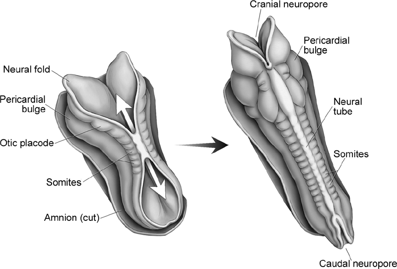
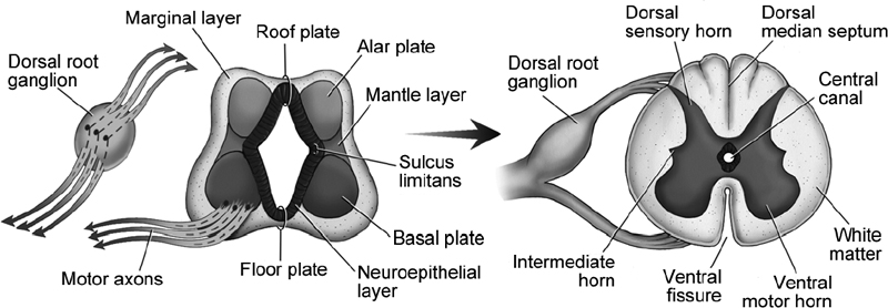
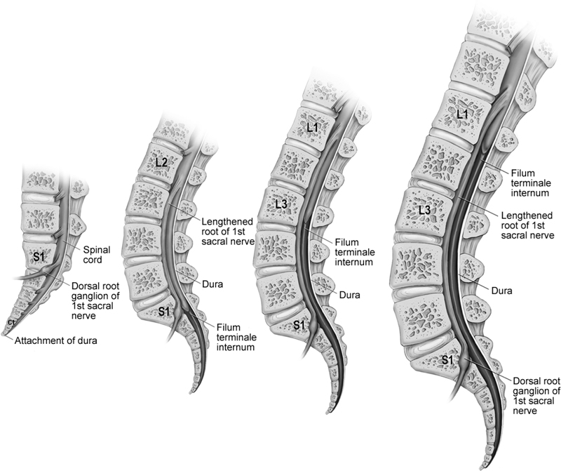
1 Embryology of the Spine I. Key Points – The developing vertebral column is formed from somites, which develop into sclerotomes. – Myotomes bridge the intervertebral spaces, allowing them to develop musculature that affords movement of the spine. – The developing sclerotomes undergo chondrification and then ossification to form each vertebral unit. – The HOX genes regulate the shape of each vertebral body.1 – Epiblast cells migrate to form the primitive groove, which in turn forms the notochord. – The anterior neuropore closes on day 25 and the posterior on day 27. – Neuroblasts form the mantle layer; the ventral portion forms the basal plates (motor) and the dorsal portion forms the alar plates (sensory). – The caudal portion of the tube undergoes retrogressive differentiation and relative ascension of the conus. II. Bony Anatomy – Paraxial mesoderm forms 42 to 44 somites. – Somites differentiate into ventromedial sclerotomes and dorsolateral dermomyotomes. – In week 4, cells of the sclerotomes move to surround the spinal cord and notochord.2 – The sclerotome can be divided into a cranial area of loosely packed cells and a caudal area of densely packed cells with a “cell-free space” in between.2 – Sclerotomes are separated by intersegmental mesenchyme and flanked by segmental nerves, myotomes, and intersegmental arteries. Sclerotomes develop into definitive vertebrae, which causes myotomes to bridge intervertebral spaces, allowing movement of the spine. – Between days 40 and 60, the process of chondrification begins and ossification follows, leading to the formation of each vertebral unit. – Cells from the caudal portion of the sclerotome migrate to the cell-free space to form the annulus fibrosus of the disc, and regressing notochord forms the nucleus pulposus. – The caudal portion of each sclerotome fuses to the cephalic portion of the adjacent sclerotome during week 6. After fusion, arteries cross vertebral bodies, with the nerves residing between them. – Fusion of adjacent sclerotomes creates the centrum, which develops into the vertebral body. – Cells adjacent to the neural tube form vertebral arches (or neural arches) that consist of the posterior elements. – In general, each vertebra develops from three primary ossification centers: one for the body and one for each half of the vertebral arch. • There are five secondary ossification centers for subaxial vertebrae, located at the superior and inferior end plates of the body, the spinous process, and the tip of each transverse process. • C1 develops from the three primary ossification centers for the left and right posterior arches and for the anterior arch. • C2 develops from five primary ossification centers: two for the body of the dens, one for the vertebral body, and one each for the left and right neural arches.3 – The shapes of different vertebral bodies are regulated by HOX genes.1 – The thoracic kyphosis is present during the fetal period, and the cervical and lumbar lordoses develop after birth. III. Neural Anatomy – By week 2, the embryo begins gastrulation and has two layers: epiblast and hypoblast. During gastrulation, epiblast cells migrate to the dorsal midline to form the primitive streak and subsequently the primitive groove. – At the end of gastrulation there are three layers: ectoderm, mesoderm, and endoderm. – At the edge of the primitive groove is a pit, the primitive node, where the notochordal process is formed by migrating epiblasts. – By day 18 the primitive groove has regressed caudally and the notochord has formed. – At three weeks’ gestation, the edges of the neural plate begin to elevate to form neural folds, and their subsequent fusion in the cervical region forms the neural tube (Fig. 1.1).2 • The anterior neuropore closes on day 25. • The posterior neuropore closes on day 27. – Neural crest cells detach from the neural folds and migrate to form glia, arachnoid, pia, melanocytes, chondrocytes, chromaffin cells, osteocytes, Schwann cells, and enteric ganglia. – The wall of the neural tube consists of neuroepithelial cells forming a pseudostratified epithelium connected by junctional complexes that differentiate into neuroblasts.2 – Neuroblasts form the mantle layer around the neuroepithelial layer that forms the gray matter of the spinal cord (Fig. 1.2).2 • The ventral mantle layer forms the basal plates (motor horn), and the dorsal mantle layer forms the alar plates (sensory horn). • At the thoracic (T1 to T12) and upper lumbar (L1 to L2) region, the intermediate horn contains sympathetic neurons of the autonomic nervous system. • The boundary between the basal and alar plates is the sulcus limitans. – The marginal layer contains nerve fibers from neuroblasts in the mantle layer that ultimately form the white matter of the spinal cord. – The caudal tube forms during canalization (days 28 to 42). – From day 43 to day 48, the ventriculus terminalis (a cystic structure at the caudal neural tube end) undergoes retrogressive differentiation, which is completed postnatally at 2 months.2 • This results in relative ascension of the conus to its final level at L1-L2, and formation of the cauda equina and filum terminale (Fig. 1.3). Fig. 1.1 Dorsal view of the human embryo during the third week of gestation. Note the somites on each side of the neural tube as it begins to fuse in the cervical region. The fused neural tube then continues to close both rostrally and caudally. Fig. 1.2 Cross-section of the developing spinal cord demonstrates how the migrating neuroblasts from the neuroepithelial layer form dorsal and ventral mantle layers. These ultimately become the gray matter of the spinal cord. In addition, note the development of the dorsal root ganglion as well as the outward growth of the motor axons. IV. Surgical Pearls – Failure of the ventriculus terminalis results in a terminal myelocystocele. This cyst is lined with ependyma and communicates with the central canal. – The sulcus limitans is the border between the sensory (dorsal) and motor (ventral) areas. – The conus ascends to its adult level, L1-L2, by 2 months of age. It is important to keep this in mind when performing a lumbar puncture on the neonate. – The notochord involutes and remains to develop the nucleus pulposus of the intervertebral disc – Complete fusion of the ossification centers of C2 does not occur until age 12. Prior to this, synchondroses between ossification centers can be mistaken for fractures. Fig. 1.3 The relative ascension of the conus and formation of the filum terminale via retrogressive differentiation. Common Clinical Questions 1. On which day does the anterior neuropore close? 2. On which day does the posterior neuropore close? 3. The shapes of various groups of vertebral bodies are governed by which set of genes?
 The tip of the dens represents a secondary ossification center.
Related posts:


Stay updated, free articles. Join our Telegram channel

Full access? Get Clinical Tree


