Enlarged Vertebral Body, Soap Bubble Expansion
Lubdha M. Shah, MD
DIFFERENTIAL DIAGNOSIS
Common
Metastases, Lytic Osseous
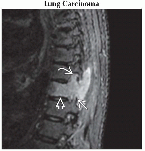
Lung Carcinoma
Thyroid Carcinoma
Renal Cell Carcinoma
Multiple Myeloma
Osteoblastoma
Giant Cell Tumor
Aneurysmal Bone Cyst
Less Common
Chordoma
Chondrosarcoma
Rare but Important
Fibrous Dysplasia
Telangiectatic Osteosarcoma
Enchondroma
Angiosarcoma
Cystic Angiomatosis
ESSENTIAL INFORMATION
Key Differential Diagnosis Issues
Zone of transition is helpful to assess aggressiveness
Multiplicity of lesions, soft tissue component, & vascularity of lesions can be helpful in narrowing the differential diagnosis
Helpful Clues for Common Diagnoses
Metastases, Lytic Osseous
Lung, thyroid, renal, breast, oro-/nasopharyngeal carcinoma
Destructive lesion involving the posterior cortex & pedicle
Intervertebral discs are spared
Location proportionate to red marrow (lumbar > thoracic > cervical)
Multiple Myeloma
Multifocal malignant proliferation of monoclonal plasma cells leads to heterogeneous T1 marrow signal
May be expansile, but vertebral compression is more common
Vertebral body more frequently involved
Pedicle involvement later than with metastases
Osteoblastoma
Ovoid expansile mass originating in the neural arch, often extend into the vertebral body
40% in spine
40% cervical, 25% lumbar, 20% thoracic, 15-20% sacrum
Florid edema (corona effect) suggests an aggressive process, attributable to prostaglandin release by the tumor
Peritumoral edema enhances avidly with gadolinium administration
Usually demonstrates more discrete bone matrix as compared to fibrous dysplasia
Bone scan demonstrates avid radionuclide uptake by the tumor
Giant Cell Tumor
Expansile, lytic lesion with narrow zone of transition
± Cortical breakthrough
Centered in vertebral body
Margin usually not sclerotic
± Residual bone trabeculae
Aneurysmal Bone Cyst
Expansile lesion may show cortical breakthrough
Shows a narrower zone of transition
Centered in posterior elements
Can be associated with fibrous dysplasia
Helpful Clues for Less Common Diagnoses
Chordoma
Midline soft tissue mass with osseous destruction
T2 hyperintense mass with multiple septa
Can involve adjacent vertebral bodies by extension across disc space
Arise from notochord remnants
Chondrosarcoma
Lytic mass ± chondroid matrix, “rings & arcs”
Cortical disruption
Extension into soft tissues
Nonenhancing areas: Hyaline cartilage, cystic mucoid tissue, necrosis
Neural arch involved more frequently than vertebral body
Helpful Clues for Rare Diagnoses
Fibrous Dysplasia
Well-defined, expansile, radiolucent lesion
Neural arch involved more frequently than the vertebral body
Spine involvement typically in polyostotic disease
Fusiform bone expansion with “ground-glass” matrix
Heterogeneous T1/T2 signal & heterogeneous enhancement
Paraspinal soft tissue extension & vertebral collapse rare
Prevalence of scoliosis in patients with polyostotic fibrous dysplasia & spinal lesions is reported between 40% and 52%
Telangiectatic Osteosarcoma
Wide zone of transition with adjacent bone
Permeative appearance & cortical disruption
Multiple fluid-fluid levels
Soft tissue mass ± mineralization
Enchondroma
Expansile, homogeneous, slightly enhancing lesion with or without calcification
Arise either from migration of hyperplasitic immature spinal cartilage outside vertebral axis
Or from metaplasia of connective tissue in contact with the spine or annulus fibrosus
Common benign cartilaginous tumour involving the acral skeleton but extremely rare in the vertebral column (2% of cases)
Angiosarcoma
Lumbar region is most commonly affected
33% in axial skeleton
Coarse trabecular/honeycomb pattern is suggestive of a vascular tumor
Cystic Angiomatosis
Lytic, well-defined, round or oval lesions within the medullary cavity
Intact cortex & variable peripheral sclerosis
Endosteal scalloping & honeycombed or latticework appearance
Discrete circular or serpentine lytic areas within bone suggest vascular channels
No periosteal reaction
SELECTED REFERENCES
1. Leet AI et al: Fibrous dysplasia in the spine: prevalence of lesions and association with scoliosis. J Bone Joint Surg Am. 86-A(3):531-7, 2004
2. Murphey MD et al: From the archives of the AFIP. Musculoskeletal angiomatous lesions: radiologic-pathologic correlation. Radiographics. 15(4):893-917, 1995
3. Kumar R et al: Expansile bone lesions of the vertebra. Radiographics. 8(4):749-69, 1988
Image Gallery
 Sagittal STIR MR shows a hyperintense lesion expanding a thoracic vertebral body
 , the articular facets , the articular facets  , & epidural space , & epidural space  . .Stay updated, free articles. Join our Telegram channel
Full access? Get Clinical Tree
 Get Clinical Tree app for offline access

|