Fig. 48.1
Fibrosarcoma. Sagittal noncontrast MRI of the pituitary (arrows) shows a spontaneously sclerosing epithelioid fibrosarcoma extending superiorly to the optic chiasm (Adapted with permission from Massier et al. [11].)

Fig. 48.2
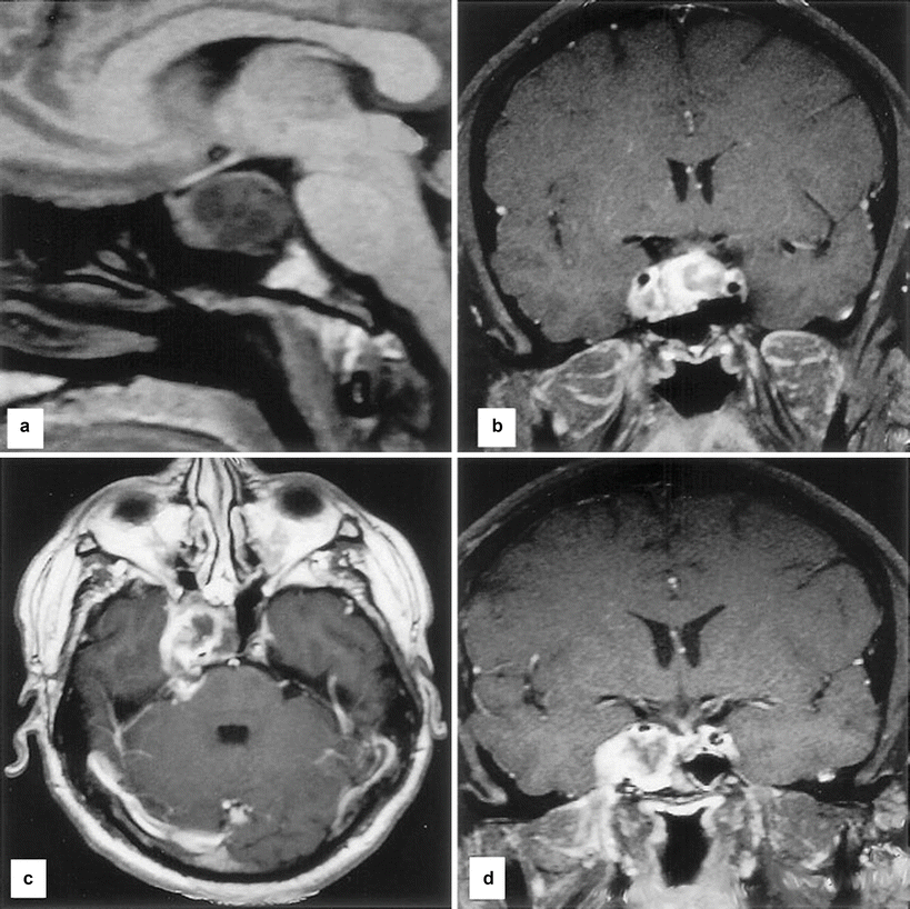
MRI of a patient with a radiation-induced sellar fibrosarcoma. (a) Sagittal MRI (T1-weighted) showing the partially cystic tumor with extension toward the optic chiasm and the brainstem. (b) Coronal MRI (T1-weighted, gadolinium-enhanced) showing inhomogeneous enhancement of the tumor with extensive intrasellar growth, suprasellar extension into the right cavernous sinus, and mild dislocation of the right internal carotid artery; there is also growth into the sphenoid sinus. (c) Axial MRI scan 5 months after radiosurgery (T1-weighted, gadolinium-enhanced), showing further tumor growth with infiltration of the pterygopalatine fossa and the lateral and medial pterygoid muscle on the right side. (d) Coronal MRI scan 5 months after radiosurgery (T1-weighted, gadolinium-enhanced), showing further infiltration of the right cavernous sinus and the right sphenoid sinus, with perineural growth along the mandibular branch of cranial nerve V (Adapted with permission from Berkmann et al. [2].)
48.3 Histopathology
The histopathological features of fibrosarcoma of the pituitary region are similar to those arising in soft tissues.
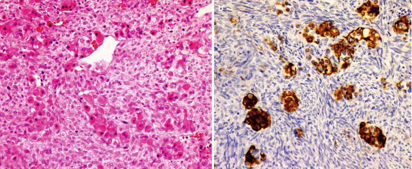
The tumors are composed of spindle-shaped cells with nuclear pleomorphism, and frequent mitoses are commonly observed (Fig. 48.3) [13].
Immunoreactivity for vimentin is seen in fibrosarcomas. These tumors lack immunostaining for S100, cytokeratin, desmin, and CD34 [5].
If the fibrosarcoma is a secondary tumor following treatment of a primary pituitary adenoma, some retained areas of pituitary adenoma pathology may be interspersed with the fibrosarcoma [5].

Fig. 48.3
Primary fibrosarcoma of the sella turcica diffusely involving the anterior pituitary. The tumor is composed of spindle cells with elongated nuclei arranged in fascicles. Note the pituitary neuroendocrine cells entrapped by the tumor, as seen by H&E (a) and highlighted by chromogranin (b)
48.4 Clinical and Surgical Management
Transsphenoidal approaches and craniotomy have been used for partial debulking. The consistency of these tumors is typically quite firm [8].
Stay updated, free articles. Join our Telegram channel

Full access? Get Clinical Tree


