Fracture, Posterior Element
Julia Crim, MD
DIFFERENTIAL DIAGNOSIS
Common
Cervical
Hyperextension Injury, Cervical
Burst Fracture, Cervical
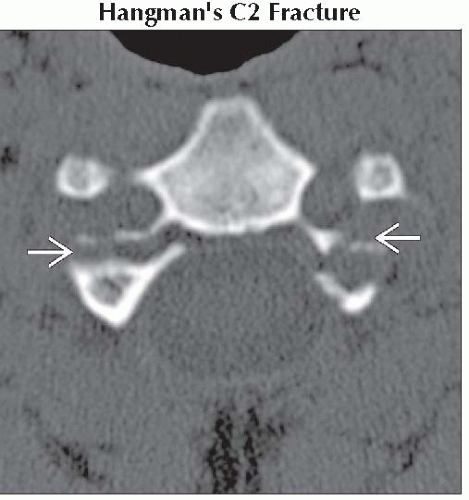
Hangman’s C2 Fracture
Lateral Flexion Injury, Cervical
Jefferson C1 Fracture
Burst Fracture, C2
Clay Shoveler’s Fracture
Hyperflexion-Rotation Injury, Cervical
Hyperflexion Injury, Cervical
Thoracic
Chance Fracture, Thoracic
Burst Thoracolumbar Fracture
Facet-Lamina Fracture, Thoracic
Lumbar
Burst Fracture, Lumbar
Spondylolysis
Facet-Posterior Fracture, Lumbar
Transverse Process Fracture
Chance Fracture, Lumbar
Pedicle Stress Fracture
All Spinal Levels
Pathologic Vertebral Fracture
Less Common
Fracture Mimics
Metastases, Lytic Osseous
Incomplete Fusion, Posterior Element
Neurogenic (Charcot) Arthropathy
Vertebral Segmentation Failure
Hypoplastic Rib, Supernumerary Rib
ESSENTIAL INFORMATION
Key Differential Diagnosis Issues
Must distinguish between isolated posterior column injury and multicolumn injury
Isolated posterior column
Due to lateral flexion or rotation, direct blow, or hyperflexion
Clay shoveler’s fracture a stable hyperflexion injury
Multiple column
Due to any injury mechanism, not necessarily unstable
Non-bony involvement of middle, anterior columns often best seen on MR
Helpful Clues for Common Diagnoses
Prevertebral soft tissues of cervical spine may be normal in isolated posterior element fracture
Burst fracture: Posterior column fracture in vertical plane
Flexion-distraction fracture: Posterior column fracture in horizontal plane
Spondylolysis easily missed on axial images; use following signs
“Double facet” sign: Spondylolysis is anterior to facet joint
“Wide canal” sign: Increased AP dimension of spinal canal
Image Gallery


