Fig. 42.1
Gangliocytoma in the presence of a prolactinoma. This sagittal T1-weighted gadolinium-enhanced MR image shows a heterogeneously enhancing mass of the sellar region with sellar expansion (Adapted with permission from Mikami et al. [14])
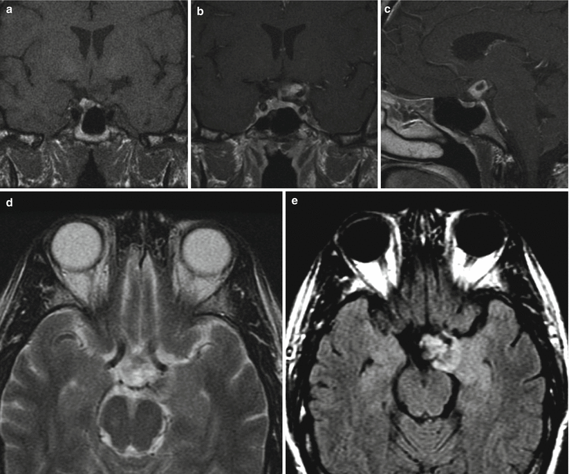
Fig. 42.2
Ganglioglioma. (a) Coronal T1-weighted precontrast MR image. (b) Coronal T1-weighted gadolinium-enhanced image. (c) Sagittal T1-weighted gadolinium-enhanced image. (d) Axial T2-weighted image. (e) Axial T2-FLAIR image. An enhancing mass is located in the left suprasellar region, contiguous with the left temporal lobe uncus. The mass mildly elevates the optic chiasm on the left and abuts the pituitary stalk and supraclinoid left internal carotid artery
42.3 Histopathology
42.3.1 Gangliocytoma
In most cases, an intimate microscopic association between gangliocytomatous elements and pituitary adenoma cells is observed. The proportion of pituitary adenoma and neuronal elements may vary widely [16, 17].
The ganglion cells are mostly large, pyramidal neurons immersed within variably prominent neuropil-like stroma.
Ganglion cells are readily identified by immunoreactivity for neuronal-associated proteins such as neurofilament proteins (NFPs) and synaptophysin and silver impregnation preparations (Fig. 42.3) [3].
Glial elements are not present, confirming the pure ganglionic derivation of these tumors.
The ganglion cells may also express hypothalamic hormones including GHRH, somatostatin, CRH, or GnRH [16].
The most frequently associated pituitary adenoma is a GH-secreting adenoma, particularly the sparsely granulated and GH/PRL cell subtypes [3, 4, 16].
These tumors have also been reported to coexist with adrenocorticotropic hormone (ACTH) cell adenomas and prolactinomas [14, 18–20].
Three major hypotheses have been considered for the histogenesis of such tumors:
1.
Displaced hypothalamic neurons in the sellar region might produce hypothalamic-releasing factors that would stimulate the proliferation of an adenoma [1].
2.
3.
Both neuronal and adenomatous components may have a common origin from progenitor/stem cells present in the adult adenohypophysis that are capable of multidirectional differentiation [21].
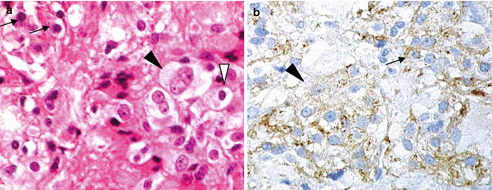
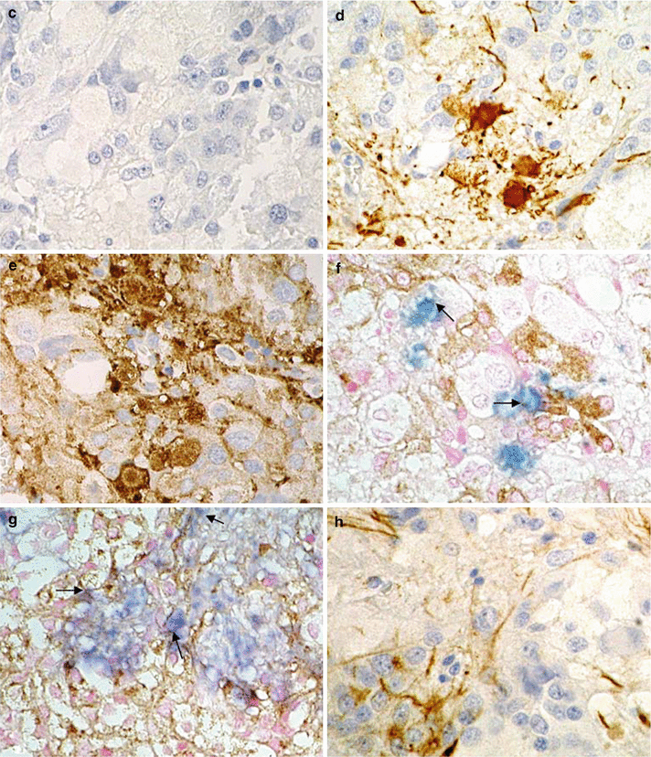
Fig. 42.3
Gangliocytoma in the presence of a prolactinoma. (a) With H&E staining, the tumor can be seen to be composed of small, round cells corresponding to the pituitary adenoma (arrows) and large ganglions (black arrowhead). Tumor cells showing intermediate features (white arrowhead) are also observed. (b) With PRL staining, adenoma cells (arrow) are immunoreactive for PRL. Ganglions are also weakly positive for PRL (black arrowhead). (c) GH staining is negative. (d) NF staining is positive in the fibrillary substrates of ganglions. (e) Synaptophysin (SYN) staining is observed in the ganglions. (f, g) Double immunostaining shows that the regional distribution of PRL (brown) and NF (blue) overlaps in some areas. (h) Cytokeratin stain CAM5.2 is strongly positive in fibrillary structures and rather weak in adenoma cells (Adapted with permission from Mikami et al. [14])
42.3.2 Ganglioglioma
Gangliogliomas of the sellar region appear to have similar histopathological features as those tumors arriving in other locations of the CNS—i.e., a variable combination of ganglionic and glial cells (Fig. 42.4).
A case of ganglioglioma was reported to demonstrate immunoreactivity for vasopressin in a patient presenting with signs of SIADH [13]. In both gangliocytomas and gangliogliomas, ultrastructural findings on electron microscopy may be useful to confirm the presence of synaptic vesicles corresponding to neuronal elements [11].
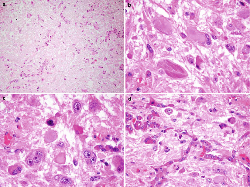
Fig. 42.4
Ganglioglioma. Microsections of the expanded asymmetric posterior lobe show accumulation of ganglion cells and scant astrocytes (a). Ganglion cells are dysmorphic (b) and feature neurofibrillary tangles (c). Note the association of ganglion cells with basophil invasion (d) (Adapted with permission from Scheithauer et al. [12])
42.4 Clinical and Surgical Management
Resection of gangliocytoma and ganglioglioma via transsphenoidal approaches and open craniotomy has been reported [23, 24].Related posts:

Stay updated, free articles. Join our Telegram channel

Full access? Get Clinical Tree


