32 Hemispheric asymmetries
The two cerebral hemispheres are asymmetrical in certain respects. Some of the asymmetries have to do with handedness, language, and complex motor activities; other, more subtle differences come under the general rubric of cognitive style. (Limbic asymmetries are described in Ch. 34.)
Handedness and Language
Handedness often determines the hemisphere that is dominant for motor control. Left hemisphere/right-hand dominance is the rule. Advances in ultrasound technology have made it possible for motor behavior in the fetus to be observed, and it has been noted that handedness is already established before birth on the basis of the preferred hand used for thumb sucking during fetal life.
The best indicator available for population estimates of handedness is the preferred hand for writing: this criterion indicates a left hemisphere dominance for motor control in about 90% of the population.
In 90% of subjects, the left hemisphere is dominant for language. In a further 7.5%, the right hemisphere is dominant in both sexes, and in the remaining 2.5%, the two hemispheres have an equal share. Although the left hemisphere is dominant in respect of both motor control and language, the two features are statistically independent: many left-handers have their language areas in the left hemisphere.
Language areas
Although several areas of the cortex, notably in the frontal lobe, are active during speech, two areas are specifically devoted to this function.
Broca’s area (Figure 32.1)
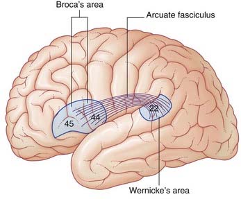
Lesions involving Broca’s area are associated with expressive aphasia (see Clinical Panel 32.1). Some workers believe that expressive aphasia requires that the lesion also includes the lower end of the precentral gyrus.
Clinical Panel 32.1 The aphasias
Aphasia is a disturbance of language function caused by a lesion of the brain. The usual cause is a stroke produced by vascular occlusion in the anterior cortical territory of the left middle cerebral artery.
Motor (anterior) aphasia
Patients having a lesion that includes Broca’s area suffer from motor aphasia. These patients have difficulty in expressing what they want to say. Speech is slow, labored, and characteristically ‘telegraphic’ in style. The important nouns and verbs are spoken but prepositions and conjunctions are omitted. The patient comprehends what other people are saying and is well aware of being unable to speak fluently. There is usually an associated agraphia (inability to express thoughts in writing).
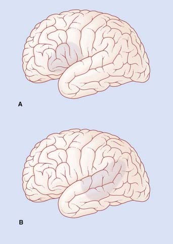
If the lesion involves a substantial amount of the cortical territory of the middle cerebral artery, there will be a motor weakness of the right lower face and right arm. Because the lips and tongue are affected on the right side, the patient will also have dysarthria (difficulty in speech articulation) in the form of slurring of certain syllables. In the example shown in Figure CP 32.1.1A, Broca’s aphasia would be associated with right-sided weakness of the lower face but not of the arm, together with some dysarthria.
Sensory (posterior) aphasia
A lesion in Wernicke’s area is accompanied by a deficit of auditory comprehension. If the lesion includes the angular gyrus, the ability to read will also be compromised. In addition to their difficulty in understanding the speech of others, these patients lose the ability to monitor their own conversation, and usually have difficulty in retrieving correct descriptive names. Speech fluency is normal but two kinds of abnormality occur in the use of nouns:
The most striking feature of Wernicke’s aphasia is that, despite garbling to the point of being unintelligible (jargon aphasia), the patient may be quite unaware of making mistakes.
In the example shown in Figure CP 32.1.1B, Wernicke’s aphasia would be associated with alexia (angular gyrus), ideomotor apraxia (supramarginal gyrus) and probably with a right upper quadrant visual deficit (lower fibers of left optic radiation in the temporal white matter).
Aprosodia
Lesions of the right hemisphere may affect speech in subtle ways. Lesions that include area 44 (corresponding to Broca’s area on the left) tend to change the patient’s speech to a dull monotone. On the other hand, lesions that involve area 22 (corresponding to Wernicke’s area) may lead to listening errors, e.g. being unable to detect inflections of speech; the patient may not know whether a particular remark is intended as a statement or as a question.
Wernicke’s area (Figure 32.1)
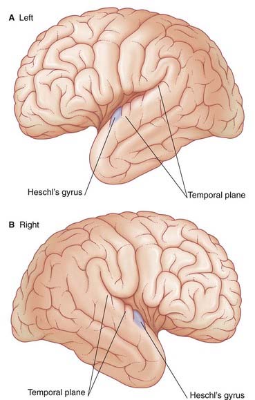
The German neurologist Karl Wernicke made extensive contributions to language processing in the late 19th century. He designated the posterior part of area 22 in the superior temporal gyrus of the left hemisphere as a sensory area concerned with understanding the spoken word. The upper surface of Wernicke’s area is called the temporal plane (Figure 32.2). The volume of cerebral cortex in the temporal plane is larger on the left side in 60% of subjects. The horizontal part of the lateral fissure is longer in consequence – a feature readily identified on MRI scans. Lesions involving Wernicke’s area in adults are associated with receptive aphasia (see Clinical Panel 32.1).
Wernicke’s area is linked to Broca’s area by association fibers of the arcuate fasciculus curving around the posterior end of the lateral fissure within the underlying white matter (Figure 32.1). The two areas are also linked through the insula.
Maldevelopment of the left temporal plane is a significant feature in cases of schizophrenia (Clinical Panel 32.4).
Right hemisphere contribution
During normal conversation there is some increase in blood flow in areas of the right hemisphere matching those of the left. These areas are believed to be concerned with melodic aspects of speech – the cadences, emphases, and nuances, collectively called prosody. Disturbances of the melodic function are called aprosodias (Clinical Panel 32.1).
Angular gyrus
The angular gyrus (area 39) belongs descriptively to the inferior parietal lobule. The left angular gyrus receives a projection from the inferior part of area 19 (the lingual gyrus, shown in Figure 2.6), and itself projects to the temporal plane. It is commonly included as a part of Wernicke’s area.
Listening to spoken words
Figure 32.3 contrasts regional increases in blood flow during PET scanning when a volunteer listens to words (‘active listening’) vs random tone sequences (‘passive listening’). As expected, tone sequences activate the primary auditory cortex (bilaterally). Wernicke’s area (left side) also becomes active, probably in screening out this non-verbal material from further processing. Area 9 in the frontal lobe is thought to be part of a supervisory, vigilance system.
During active listening to words, areas 21 (middle temporal lobe), 37 (posteroinferior temporal lobe), and 39 (angular gyrus) all participate in auditory word processing. Area 39 identifies phonemes. Areas 21 and 37 identify words in the sound sequence and tap into lexicons (dictionaries) stored in memory in a search for meaning – a process called semantic retrieval.
Activity in the left dorsolateral prefrontal cortex (DLPFC) expands to include area 46. Engagement of Broca’s area is thought to signify ‘subvocal articulation’ of words heard (see Neuroanatomy of reading, later).
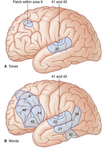
When listening to one’s own voice, the areas of the temporal lobe identified above become active. An important function being served here is meta-analysis (post hoc analysis) of speech, whereby ‘slips of the tongue’ can be identified. Speech meta-analysis is singularly lacking in cases of receptive aphasia (Clinical Panel 32.1).
Modular organization of language
In alert subjects, electrical studies of the cortex exposed during neurosurgical procedures indicate the presence of a vast cortical mosaic for language. The mosaic of modules extends along the entire length of the frontoparietal operculum above the lateral sulcus and of the temporal operculum below the sulcus. The frontoparietal operculum is predominantly concerned with the motor functions of speaking and writing, and the temporal operculum with the sensory functions of hearing and reading.

Stay updated, free articles. Join our Telegram channel

Full access? Get Clinical Tree

