Figure 53-1 iThe extensive bleeding into sciatic nerve (A) and its multifocal distribution at different proximal to distal levels of a freshly isolated fascicle of sciatic nerve before fixation (B) is easily recognized. In a hematoxylin and eosin–stained paraffin section of sciatic nerve, fresh blood, and degradation products of hemoglobin (bilirubin and biliverdin, yellow-brown) are especially apparent in perineurium and epineurium (C). The iron (arrows) from degraded hemoglobin is visualized using the Turnbull blue stain (D).
(Reprinted with permission from Moon JS, Dyck PJB, Engelstad JK, Witt LV, Letendre L, Dyck PJ. Hemorrhagic polyneuropathy in idiopathic thrombocytopenia purpura. J Peripher Nerv Syst 2007;12:286-9.)
In paraffin sections, the focal hemorrhagic lesions contained not only large amounts of fresh blood but also degradation products of hemoglobin such as bilirubin, biliverdin, and hemosiderin, both free and in macrophages (see Fig. 53-1C and D). The perineurial and subperineurial bleeding displaced endoneurial contents (Fig. 53-2).
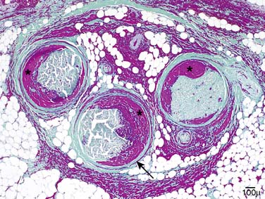
Figure 53-2 The massive epineurial (the violet-red staining around and among fascicles), endoneurial (the contents of fascicles as marked, asterisks) and perineurial (arrow) bleeding is shown from a section of sciatic nerve from our patient (Masson’s trichrome stain).
(Reprinted with permission from Moon JS, Dyck PJB, Engelstad JK, Witt LV, Letendre L, Dyck PJ. Hemorrhagic polyneuropathy in idiopathic thrombocytopenia purpura. J Peripher Nerv Syst 2007;12:286-9.)
In order to further test the hypothesis that axonal degeneration is secondary to hemorrhage, individual fascicles were isolated and sectioned from serial blocks along the length of nerves. Both gross and microscopic studies were done. Among 32 fascicles, 22 had a little or no bleeding, whereas 10 fascicles had patchy and sometimes gross bleeding. We studied teased nerve fibers from 5 fascicles with bleeding and 3 fascicles without bleeding. Almost all (96-98%) of teased myelinated fibers (MF) from all fascicles were undergoing acute axonal degeneration both at the rootlet and the sciatic nerve levels. Axonal degeneration occurred in both fascicles with and without intrafascicular bleeding.
On semithin epoxy sections and electron microscopy, axonal degeneration was prominent. Furthermore, there were frequent MF with swollen dark axons, containing many organelles, vesicles, mitochondria, tubules, dense organelles and breakdown products (Fig. 53-3). Other features observed were erythrocytes within degenerating/regenerating Schwann cell tubes that also contained Schwann cells and MF breakdown products (see Fig. 53-3). We did not find lymphomatous infiltration into nerves.
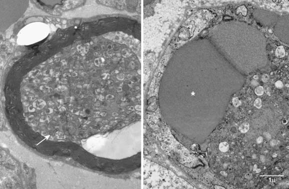
Figure 53-3 Electron micrographs of a degenerating myelinated fiber (left) and a degenerating/regenerating Schwann cell tube (right) of sciatic nerve of our patient. The figure on the left shows accumulated organelles (arrow), which is thought to be due to alterations of rapid axonal flow, perhaps from ischemia. The degenerating/regenerating Schwann cell tube (Band of Büngner) on the right contains two erythrocytes (asterisk). We have not previously encountered erythrocytes in such degenerating/regenerating Schwann cell tubes.
(Reprinted with permission from Moon JS, Dyck PJB, Engelstad JK, Witt LV, Letendre L, Dyck PJ: Hemorrhagic polyneuropathy in idiopathic thrombocytopenia purpura. J Peripher Nerv Syst 2007;12:286-9.)
Related posts:
 Malignant Peripheral Nerve Sheath Tumor
Disseminated Sporotrichosis with Multiple Granulomatous Mononeuropathies
Late Sporadic CMT4C—A New KIAA1985 Mutation
Late-Onset Transthyretin Val30Met Familial Amyloid Polyneuropathy Unrelated to Endemic Foci
A Weak, and Numb Patient with Tremor—Antimyelin-Associated Glycoprotein Polyneuropathy
Copper Deficiency Myeloneuropathy
Malignant Peripheral Nerve Sheath Tumor
Disseminated Sporotrichosis with Multiple Granulomatous Mononeuropathies
Late Sporadic CMT4C—A New KIAA1985 Mutation
Late-Onset Transthyretin Val30Met Familial Amyloid Polyneuropathy Unrelated to Endemic Foci
A Weak, and Numb Patient with Tremor—Antimyelin-Associated Glycoprotein Polyneuropathy
Copper Deficiency Myeloneuropathy
Stay updated, free articles. Join our Telegram channel

Full access? Get Clinical Tree


