Historical Aspects of EEG
Ernst Niedermeyer
Donald L. Schomer
DISCOVERY OF ELECTRICAL PHENOMENA
Thales from Miletos has been credited with the discovery of static electricity produced by friction (rubbing fur or glass with silk). He was one of the pre-Socratic “natural philosophers” of Greece (around 620-550 BC) and considered water the origin of all things. Thus, friction was recognized as the generator of a phenomenon that derived its name from the Greek work “electron,” which stands for amber. This discovery fell into a dormant stage for more than two millennia.
Around 1600, William Gilbert began to study the electrical properties of various substances, and Otto von Guericke (1602-1686) invented the friction machine to create electrical fields. This machine eventually found its way into doctors’ offices and even university hospitals. Its electrical field would make a patient’s hair stand up, creating a strong impression on a psychologically gullible patient. These friction machines now ornament high school laboratories and technical museums. In the 17th and 18th centuries, the friction machine taught invaluable lessons on attraction and repulsion of charged bodies, on conductors and nonconductors, and on the rather questionable dualism of positive and negative electricity.
A new and very important piece of electrical equipment entered the scene in 1746 when the Leyden jar was introduced by Pieter van Musschenbroek (following the earlier work of Ewald von Kleist). This invention resulted in the storage of electricity, and its upshot, the condenser or capacitor, turned into an indispensable part of modern electronics. Benjamin Franklin’s bold experiment caught electrical discharges of a thunderstorm in a Leyden jar.
What the friction machine could generate, the Leyden jar could store. Its sudden discharge was used in many experiments (O’Leary and Goldring, 1976).
The role of static electricity in medicine appeared to be forgotten for about 150 years and became resurrected with the introduction of the defibrillating cardioversion by William B. Kouwenhoven and his coworkers in the 1950s and 1960s; this approach may hold promises for cerebral applications (Niedermeyer, 2003a).
A serious scientific controversy developed in Italy between Luigi Galvani (1737-1798), a professor at the University of Bologna, and Alessandro Volta (1745-1832) in the wake of Galvani’s discovery of frog leg contractions within an electrical circuit and especially in the presence of a thunderstorm (1780). Volta doubted the biologic nature of the contraction (animal electricity) and placed the emphasis on physics—on his “pile,” the first battery (around 1800). This bimetallic pile was a generator capable of producing a steady flow of electricity. Volta’s view more or less prevailed in this hotly debated argument. The laws governing flowing electricity were soon discovered by Georg Ohm in 1827.
Nevertheless, Galvani’s belief in “animal electricity” was not lost with other discarded false ideas. There still remained the nagging question of an active electrical contribution of animal muscle tissue.
BEGINNINGS OF ELECTROPHYSIOLOGY
The introduction of the galvanometer has been associated chiefly with the name of Nobili in Florence; this instrument was refined in 1858 by William Thompson (Lord Kelvin) in England (O’Leary and Goldring, 1976). These galvanometers would faithfully demonstrate continuous electrical currents and their variations in intensity but failed in the detection of instantaneous electrical phenomena.
Carlo Matteucci (1811-1868) in Bologna and Emil Du Bois-Reymond (1818-1896) in Berlin became the major proponents of an electrophysiologically based physiology of the nervous system. (The French name of Du Bois-Reymond indicates the Huguenot origin of this Prussian investigator.) Du Bois-Reymond coined the term negative variation for a phenomenon occurring during muscle contraction when the galvanometer indicated an unexpected decrease in current intensity (O’Leary and Goldring, 1976). This term was later resurrected in earliest electroencephalogram (EEG) research (Caton, 1875) and with the discovery of the “contingent negative variation” (Walter, 1964).
Hermann von Helmholtz (1821-1894) accurately measured the velocity of nerve conduction, which had been vastly overestimated up to that time. The electrodes used in physiologic research were improved and made nonpolarizable (Du Bois-Reymond). The concept of “action current” was introduced by L. Hermann (1834-1919) and thus clarified Du Bois-Reymond’s negative variations found during muscle contraction. Julius Bernstein (1839-1917) proposed a membrane theory of nerve tissue, which ultimately was elucidated as late as 1939 and the following years by A. L. Hodgkin and A. F. Huxley in England. Against this background of strongly evolving electrophysiology of the nervous system, the first observation of EEG-like electrical brain activity took place.
CATON: THE FIRST ATTEMPT AT THE ELECTRICAL ACTIVITY OF THE BRAIN
Richard Caton (1842-1926) (Fig. 1.1) was a physician practicing in Liverpool who became deeply interested in electrophysiologic phenomena and eventually received a grant from the British Medical Association to explore electrical phenomena of the exposed cerebral hemispheres of rabbits and monkeys. According to Brazier (1961), Caton presented his findings to the association on August 24, 1875, and a very short report of 20 lines subsequently appeared in the British Medical Journal. A more detailed report was presented in the same journal in 1877 on experiments of more than 40 rabbits, cats, and monkeys, the rabbit having been principally employed.
Caton used a galvanometer. A beam of light was thrown on the mirror of the galvanometer and reflected on a large scale placed on the wall. With this type of visualization, Caton found that “feeble currents of varying direction pass through the multiplier when the electrodes are placed on two points of the external surface, or one electrode on the grey matter, and one on the surface of the skull.” This sentence is regarded as indicating the birth of the electrophysiologram because one can assume that EEG phenomena made the needle move from one direction to the other. (The suffix “gram” naturally is out of place since “graphein” means “to write” and there was no written recording.) Even though artifacts could have played a major role, Caton deserves credit for the discovery of the fluctuating potentials that constitute the EEG.
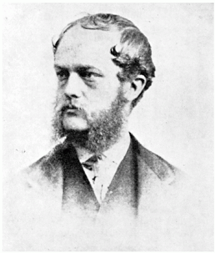 Figure 1.1 Richard Caton at the time of his work on the electrical activity of the brain. (From Brazier MAB. A History of the Electrical Activity of the Brain. The First Half-Century. London: Pitman; 1961, with permission from Macmillan.) |
Caton also described a few more interesting observations. He noted that the external surface of the gray matter was positive in relation to deep structures of the cerebrum. He also noted that the electric currents of the cerebrum appeared to have a relation to underlying function: “When any part of the grey matter is in a state of functional activity, its electric current usually exhibits negative variation.” Thus, Caton has also been credited with pioneer work on evoked potential. Furthermore, the difference in polarity found between cortical surface and deeper areas could be interpreted as the discovery of the “steady potential” (“DC potential”), but it might be wise to refrain from such statements that cannot be fully supported by the evidence. With regard to the fluctuations, Geddes (1987) pointed out that Caton’s galvanometer had a very limited frequency response range from 0 to 6 Hz.
Caton found some measure of success and recognition with this work and held the chair of physiology at the University College of Liverpool from 1884 to 1891, when he resigned from this post. Later he became dean of the medical faculty and, in 1907, mayor (Lord Mayor) of Liverpool. The electrical activity of the brain did not occupy a predominant position in his further endeavors. Even though Caton became an EEG research dropout, his bold work will always remain a milestone in the history of the electrical activity of the brain. (More information on Caton’s life and work is found in Mary Brazier’s (1961) fine account.)
EASTERN EUROPEAN STUDIES OF ELECTRICAL BRAIN ACTIVITY
The time was ripe for further studies of electrical phenomena of the cerebrum. Concurrent with Caton’s epochal work of 1875, physiologists of Eastern Europe began to demonstrate their independent observations and discoveries concerning the brain and its electrical activity. Another discovery of the 1870s had an incomparably greater impact on the neuroscientific world than Caton’s demonstration of electrical activity of the brain. The capability of the human cerebral cortex to be electrically stimulated was discovered by G. Fritsch (1838-1927) and Julius Eduard Hitzig (1838-1907) in a joint study in 1870. According to O’Leary and Goldring (1976), an unusual observation had prompted Fritsch in his work: he had observed contralateral muscle contractions during dressing of an open brain wound in the Prussian-Danish War of 1864. The work of Fritsch and Hitzig was furthered by D. Ferrier and G. F. Yeo in 1880, who performed electrical stimulations of the cerebrum in apes and also in a patient who was operated on for a brain tumor. The repercussions of the stimulation studies were considerable since many investigators of that time held the view that the entire cerebrum is a homogeneous organ that harbors mental functions.
The response of the cortex to electrical stimulation probably was a special incentive for the study of its spontaneous electrical phenomena. This incentive was particularly strong in Eastern Europe, that is, in laboratories of Russian and Polish universities. (In spite of the important historical ethnic and national differences, the fact cannot be ignored that most of Poland was part of the Czarist Russian Empire throughout the 19th century.)
Vasili Yakovlevich Danilevsky (1852-1939) was only 25 years old when he finished his thesis entitled “Investigations into the Physiology of the Brain” (Danilevsky, 1877), written at the University of Kharkov. This work was based on electrical stimulation as well as on spontaneous electrical activity in the brains of animals. Thus, Danilevsky walked in Caton’s footsteps; in 1891, he gave full credit to Caton’s priority. Mary Brazier (1961) comments on the disappointment of Danilevsky who saw his high hopes unfulfilled as far as the spontaneous electrical activity of the brain was concerned; he had expected better correlation with psychic and emotional processes. He remained deeply involved in brain physiology and published an extensive textbook of human physiology in 1915. He was not the only EEG researcher with shattered hopes in the field of psychophysiology.
The life and work of Adolf Beck (1863-1939) have been described in great detail by Brazier (1961). Beck worked in Kraków as well as in Lwow (the Polish province of Galicia, at that time a part of the Austrian-Hungarian monarchy). With nonpolarizable electrodes, Beck investigated the spontaneous electrical activity of the brain in rabbits and dogs. He observed the disappearance of rhythmical oscillations when the eyes were stimulated with light and thus became a forebear of Berger’s discovery of alpha blocking. His work became widely known due to its publication in the Centralblatt.
To present a chronologic account of the events, let us leave Eastern Europe for a moment, but not Vienna. In 1883, Ernst Fleischl von Marxow (1846-1891) deposited a sealed letter at the Imperial Academy of Sciences in Vienna that contained observations on cerebral electrical activity recorded over the visual cortex in various species of animals. He did not observe oscillatory activity. He claimed priority when Beck in 1890 published his data but was not aware of earlier work done by Caton and Danilevsky. The oddity of this episode is underscored by more recent historical accounts (Brazier, 1961; O’Leary and Goldring, 1976) that indicate that Fleischl von Marxow’s work was not of first-rate quality. This does not detract from the renaissance-man versatility of this Austrian physiologist, who was well versed in linguistics (even Sanskrit), swimming, hunting, and mountain climbing.
Exciting new studies were being conducted in Eastern European universities. Napoleon Cybulski (1854-1919), who was Beck’s teacher in Kraków and an internationally renowned leader in general physiology, presented experimental electroencephalographic studies in graphical form by using a galvanometer with a photographic attachment. He provided EEG evidence of an epileptic seizure in the dog caused by electrical stimulation.
Two Russian physiologists made further studies along these lines: Pavel Yurevich Kaufman (1877-1951) and Vladimir Vladimirovich Pravdich-Neminsky (1879-1952). Prior to the discussion of their work, a few words must be said about technological developments. The d’Arsonval galvanometer featured a mirror mounted on a movable coil; light focused on the mirror was deflected when a current passed the coil. The capillary electrometer was introduced by G. Lippmann and H. J. Marey (for further details, see O’Leary and Goldring (1976)). One of the most important advances was Willem Einthoven’s introduction of the string galvanometer in 1903, a very sensitive instrument that required photographic recording and became the standard instrumentation for electrocardiography at the turn of the century.
Kaufman’s work and life are portrayed in Brazier’s (1961) historical account. Kaufman expressed the view that an epileptic attack would have to be associated with abnormal electrical discharges, and he studied the effects of cortical electrical stimulation. With World War I, he took the name of Rostoutsev and worked mainly at the University of Baku.
Pravdich-Neminsky began recording electrical brain activity of animals in 1912 with the string galvanometer. As Brazier (1961) has pointed out, his recordings, published in 1912, were the first pictorial demonstration of EEG and appeared 2 years earlier than Cybulski’s tracings. Pravdich-Neminsky recorded the EEG from the brain, the dura, or the intact skull of the dog. He described a 12 to 14/sec rhythm under normal conditions and marked slowing under asphyxia. Furthermore, he coined the term electrocerebrogram (Fig. 1.2).
The achievements of the Eastern European neuroscientists during those 50 years preceding the outbreak of World War I fill us with awe and clearly demonstrate their special talent for electrophysiologic neurophysiology. Limiting discussion to EEG history can show only the tip of the iceberg. To assess the true strength of their neuroscientific institutions, one must mention investigators in somewhat related electrophysiologic areas. Ivan Michailovich Sechenov (1829-1905) appears to be founder of this powerful school of eminent neurophysiologists. He studied the electrical activity of the spinal cord and oblongata in the frog and was a predecessor of Pavlovian thought. Nikolai Yevgenevich Wedensky followed Sechenov as the chair and professor of physiology at St. Petersburg (known for the concept of Wedensky inhibition). Vladimir Efimovich Larionov, also working in St. Petersburg, conducted beautiful studies of the auditory cortex in the dog.
The greatest Russian neuroscientist was also the most eminent clinical neurologist of his country: Vladimir Mikhailovich Bechterev (also “Bekhterev”) (1857-1927). He occupied the chair of psychiatry in St. Petersburg, which included the field of clinical neurology. He was a disciple of Du Bois-Reymond, Paul Emil Flechsig, and Wilhelm Wundt, and also worked at Charcot’s clinic in Paris. The influence of Wundt prompted Bechterev’s associative reflexology (I treasure his work “Allgemeine Grundlagen der Reflexologie des Menschen,” Deuticke, Leipzig, 1926, even though I can hardly agree with his “objective study of personality”). He combined his clinical work and private practice with tireless psychophysiologic methods. A photograph with one of his two assistants shows Bechterev wearing a thick winter coat in his institute in Leningrad, giving testimony to the icy cold in the unheated laboratory. “Functional anatomy of the brain, experimental psychology and clinical neurology were the three fields in which Bekhterev carved out a place for himself” (Yakovlev, 1953).
The Soviet regime had to choose between Bechterev and Ivan Petrovich Pavlov (1849-1936), a physiologist who had won the Nobel Prize in 1904 for his early work on conditioned reflexes. Pavlov was the choice, and the magic of conditioned
reflex overshadowed all Soviet neurophysiology by highest decree (even though Pavlov himself was highly critical of the regime). The Pavlovian concept was closer to the ideology of dialectic materialism, and this maxim with all its intolerant dogmatism outlasted Pavlov’s death by two decades. This ideopolitically governed form of neuroscience stifled all progress of customary neurophysiology. The world leadership in EEG and related fields quickly crumbled, accompanied by a terrifying decline of a dogmatically governed neurophysiology.
reflex overshadowed all Soviet neurophysiology by highest decree (even though Pavlov himself was highly critical of the regime). The Pavlovian concept was closer to the ideology of dialectic materialism, and this maxim with all its intolerant dogmatism outlasted Pavlov’s death by two decades. This ideopolitically governed form of neuroscience stifled all progress of customary neurophysiology. The world leadership in EEG and related fields quickly crumbled, accompanied by a terrifying decline of a dogmatically governed neurophysiology.
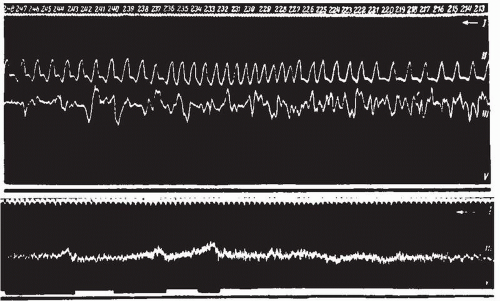 Figure 1.2 The first photographs to be published of electroencephalograms. In the upper record Neminsky shows (in the third trace) the brain potentials of a curarized dog with the pulsations from an artery in the brain recorded above them. In the lower record the sciatic nerve is being stimulated from time to time, and the decrease in activity noted by Neminsky can be seen. The record reads from right to left, line I being a time marker in one fifth of a second, line III the galvanometer string, and line V the signal for stimulation. (From Pravdich-Neminsky VV. Ein Versuch der Registrierung der elektrischen Gehirnerscheinungen. Zbl. Physiol. 1913;27:951-960, with permission from Dr. Mary Brazier and Macmillan.) |
DEVELOPMENTS IN WESTERN AND CENTRAL EUROPE
Electroencephalographic research was in a dormant state in Western and Central Europe while it was flourishing in Eastern European countries. This is quite amazing because neurophysiology in general was healthy and well outside Russia and her neighbors, but the ancestral lineage from Galvani to Du Bois-Reymond and Caton broke off and the neurophysiological field was watered by rivulets of different orientation. Thus, the work of Fleischl von Marxow lies like an erratic patch in the vast field.
The Western neurophysiologists followed attentively the work of their neuroanatomical confreres and the great controversy between network theories (the nerve cells forming a feltlike net) and the neuron theory (the neuron representing a unit). The net theory was supported by Joseph von Gerlach and by Camillo Golgi, the discoverer of the silver chromate staining method, but the neuron theory with its great principal proponent Santiago Ramón y Cajal (1852-1934) proved to be victorious in this struggle despite further attempts of new generations of “reticularist” believers in a continuous network of nerve cells.
In a similar manner, cerebral localizationists struggled against antilocalizationists. In Germany, Friedrich Leopold Goltz (1834-1902) removed the cerebral hemispheres in the “dog without cerebrum” living in a state of extreme lethargy and mental inertia (Goltz, 1888). H. Rothmann (1923) performed similar investigations. This type of research was aimed at the working of the cerebral hemispheres as a whole and deemphasized the aspects of cortical localization. As it was pointed out previously, the cerebral stimulation studies of Fritsch, Hitzig, Ferrier, and Yeo initiated a new era of interest in cortical localization.
In this period—the last third of the 19th and the dawning of the 20th century—the work of Charles Scott Sherrington (1857-1952), performed in Liverpool and Oxford, became most influential in the development of a modern Western type of reflexology. The Integrative Action of the Nervous System (Sherrington, 1906) was based on a series of lectures held at Yale University in 1904. The scope of this work reaches from reflexology to decerebrate rigidity, from motor cortex to sensory function, while the issue of mental function is being skirted with the modesty of a truly great neuroscientist. Inhibition is one of the great Sherringtonian discoveries. Even Ramón y Cajal’s net of independent synaptically connected neurons stood solely in the service of neural excitation. (Incidentally, the term synapse was introduced by Sherrington.)
It is unfortunate that this greatest master of neurophysiology stood miles away from electrophysiologic thought. His work was based chiefly on ablation techniques. He may hardly ever have given a thought to EEG methods even though he lived a full active life. His disciples—to name only Edward Liddell and Derek Ernest Denny-Brown—held similar views, while in Cambridge electrically oriented neurophysiology found its greatest proponent in Edgar Douglas Adrian (1889-1977), whom we discuss later in his relationship to Hans Berger’s work.
HANS BERGER AND THE HUMAN ELECTROENCEPHALOGRAM
Hans Berger (1873-1941) (Fig. 1.3), the discoverer of the human EEG, was a neuropsychiatrist. What neuropsychiatry really meant in those years is poorly understood nowadays. Neurology and psychiatry formed one specialty, one discipline, in Germany, Austria, and a considerable number of other countries. Neuropsychiatric departments at university hospitals and other institutions consisted of neurologic and psychiatric floors; medical specialty training meant rotation from one discipline to the other, and a the head of one department was supposed to be a master of both disciplines. Pure neurology was just beginning to emerge as a special discipline in the Germanspeaking countries (with the work of Wilhelm Erb (1840-1921), Max Nonne (1861-1959), and the incomparable Otfrid Foerster who, like Hans Berger, lived from 1873 to 1941).
Berger was not a leader, neither in neurology nor in psychiatry. Without his pioneering EEG work, his name would have been forgotten. Biographic sketches (especially Kolle, 1956) portray Berger as an extremely meticulous and conscientious person, somewhat aloof in his contact with his patients, a very strict and authoritarian department head, and an “anima candida” (a pure soul), a hard-working professor without any interest in faculty schemes and diatribes. He hardly ever attended the annual meetings of the German neuropsychiatric society. His electroencephalographic work was carried out in a small and very primitive laboratory. His first scientific interest aimed at the cerebral circulation; plethysmographic methods were used in patients with skull defects. From 1902 to 1910, he studied the electrical activity of the cerebrum in the dog with a capillary electrometer after Lippmann, but the results were disappointing. Naturally, Berger was aware of the scanty pertinent literature from Caton to Cybulski and Pravdieh-Neminsky. His studies of the human EEG started in 1920; the introduction to Gloor’s authoritative translation of Berger’s work contains a plethora of interesting details (Gloor, 1969).
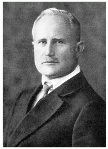 Figure 1.3 Hans Berger. (From Kolle K. Hans Berger. In: Kolle K, ed. Grosse NervenŠrzte. Vol. 1. Stuttgart: Thieme; 1956: 1-6.) |
Every electroencephalographer should be familiar with Berger’s work, an undertaking that has been greatly facilitated by Gloor’s English translation. It is true that the original German text is cumbersome and does not make easy reading, which is probably a reason for the very slow acceptance of Berger’s work. Those 14 reports bear the same title: “On the Electroencephalogram of Man.” Surely, a more attractive title would have helped somewhat. Berger’s humanistic educational background becomes obvious in the rejection of the term electrocerebrogram of Pravdich-Neminsky for strictly linguistic reasons: the “ugly” mixture of Greek (“electro,” “gram”) and Latin (“cerebro”) fragments. What Berger proposed in German was the term Elektrenkephalogram (sic) since the root enkephalo from the Greek is linguistically more correct than encephalo.
Before Berger’s work as such is brought into focus, we must discuss his electrophysiologic instrumentation. He used a string galvanometer starting in 1910—first with the Einthoven type, later with the smaller Edelmann model, and after 1924 with the larger Edelmann model. In 1926, Berger started to use the more powerful Siemens double-coil galvanometer (attaining a
sensitivity of 130 µV/cm; Grass, 1984). With this instrument and the use of nonpolarizable pad electrodes, Berger recorded the human EEG tracings shown in his first report of 1929. The records were made on photographic paper with recordings from 1 to 3 minutes’ duration. Berger used a bipolar recording technique with fronto-occipital leads for his one-channel EEG tracings along with simultaneous electrocardiogram (ECG) recording and a time marker. In 1932, he received an oscillograph from Siemens but was unable to obtain further amplifiers with oscillographs in order to obtain multichannel recordings.
sensitivity of 130 µV/cm; Grass, 1984). With this instrument and the use of nonpolarizable pad electrodes, Berger recorded the human EEG tracings shown in his first report of 1929. The records were made on photographic paper with recordings from 1 to 3 minutes’ duration. Berger used a bipolar recording technique with fronto-occipital leads for his one-channel EEG tracings along with simultaneous electrocardiogram (ECG) recording and a time marker. In 1932, he received an oscillograph from Siemens but was unable to obtain further amplifiers with oscillographs in order to obtain multichannel recordings.
Studies of the human EEG began in 1924. Appointments were made for several patients with large skull bone defects (there was no scarcity of such patients in post-World War I Germany). On July 6, 1924, the small Edelmann string galvanometer showed oscillations presumably coming from the underlying brain. In 1925, Berger recognized that skull defects were not necessarily advantageous to obtaining a recording because of thickening of dura, postoperative adhesions, etc., and he found that recordings could be made just as well (or even better) through the intact skull and scalp. Between 1926 and 1929 Berger obtained good records with alpha waves; the double-coil galvanometer was crucial for these observations. The data were often uncertain and, in 1928, Berger was beset with doubts concerning the authenticity of his observations (according to his diary entries, very impressively demonstrated by Jung, 1963).
The first report of 1929 features the alpha rhythm and the alpha blocking response (naturally along with a description of the smaller beta waves). Chlorinated silver needle electrodes, platinum wires, and zinc-plated steel needles were used in those years (Fig. 1.4). The bold first report of 1929 produced no “waves” until a confirming report came from Adrian in Cambridge (Adrian and Matthews, 1934). Throughout the 1930s, Berger’s reports on the human EEG contained veritable gems: studies of fluctuation of consciousness, first EEG recordings of sleep (the first recording of spindles), the effect of hypoxia on the human brain, a variety of diffuse and localized brain disorders, and even an inkling of epileptic discharges. Eventually, Berger was invited to an international congress of psychologists in Paris in 1937 and to Bologna where the bicentennial of Galvani’s birthday was celebrated (also 1937). His relationship to the Nazi regime was not good, and Berger was most unceremoniously made a professor emeritus at earliest convenience, in 1938. This was indeed a hard blow to his plans for further electroencephalographic studies and, in the wake of a flu-like disease, he evidently developed a severe endogenous depression, which remained undiagnosed. He ended his life by suicide on June 1, 1941 at the age of 68. External factors may have contributed to his depression: in addition to his forced retirement at 65 (a few additional “years of honor” were granted to most retiring directors of university institutions), he also felt challenged by a group of independent EEG workers at the Institute of Brain Research at Berlin-Buch. This group was led by A. E. Kornmüller and produced excellent experimental EEG work, which is discussed later. Kornmüller might have had the better connections to the government institutions in Berlin, and the highly sensitive and often insecure Berger was afraid that his discovery was being taken away from him by his more aggressive colleagues in Berlin-Buch.
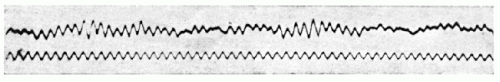 Figure 1.4 The first recorded electroencephalogram of a human. The lower line is a 10 cycles/sec sine wave for use as a time marker. The upper line is the recording from Berger’s young son made in 1925. (From Berger H. Über das Elektrenkephalogramm des Menschen. 1st report. Arch Psychiat Nervenkr. 1929;87:527-570, with permission from Dr. Mary Brazier and Macmillan.) |
Berger was a very complex person and investigator. It was pointed out previously that he did not excel clinically, neither as psychiatrist nor as neurologist (even though he was very interested in cerebral localization and particularly in the localization of brain tumors). Berger also developed very unscientific ideas about the nature of the EEG, even though he was a meticulous scientist in his EEG work. The driving force in all his research work was the quest for the nature of the all-powerful force of mental energy (“psychische Energie”). An early personal experience convinced him that such a mental energy—even capable of transmitting thoughts and emotions from person to person—does exist. According to Berger’s concept, influenced by the Danish physiologist Alfred Lehmann in 1901, mental energy is thought to be a partial product of metabolic energies (warmth and electricity being the other two products). This concept gives the EEG waves the eerie character of messengers within the mental activities, even as messengers from person to person.
Friedrich Rueckert (1788-1866), a great German poet of the Romantic period, was Berger’s maternal grandfather. Behind the strict directorial facade of Berger was the gentle soul of a highly vulnerable man. It was the psychophysiologist Berger who searched for the correlate of mental energy and, on this voyage, he found the human EEG. It was one of those “Columbus syndromes”: that a discovery is made as a by-product of a search for a different goal. Jung (1963) noted that Berger pursued his goal with the extremely powerful energy of a dilettante who had found a concept. Specialists, like the excellent physiologist (and electrophysiologist) Wilhelm Biedermann, in Jena were convinced that Berger’s dilettantism would lead nowhere, but it was the dilettante and not the seasoned specialist who emerged victorious. Even though the EEG is not exactly what Berger assumed, his contribution was the greatest in the history of electroencephalography.
BERGER’S CONTEMPORARIES
The Berliner Group
The Institute of Brain Research (Hirnforschungs-Institut) in Berlin-Bush harbored a group of ambitious and energetic investigators in various neurosciences. Oskar Vogt (1870-1959), one of the great neuroanatomists and neuropathologists of his time and a remarkably independent thinker with a wide intellectual horizon, was the director of the institute. In 1936 he lost his “directorship for lifetime” when the Nazi government became aware of Vogt’s activities at a similar institute in Moscow and his reluctance to get rid of Jewish coworkers (Hassler, 1959).
The Berliner Institute (a section of the Kaiser-Wilhelm Institutes) was composed of a variety of departments. In this context, the Department of Physiology under M. H. Fischer and the Department of Electrophysiology under A. E. Kornmüller must be singled out. These departments enjoyed the collaboration of an outstanding physicist and electronic engineer, J. F. Toennies (1902-1970), a personal friend of Oskar Vogt (both coming from the town of Husum in Holstein).
Toennies built the first ink-writing biologic amplifier for the recording of brain potentials. While in New York as a fellow of the Rockefeller Foundation in 1932, he designed the differential amplifier—the still all-important principle of EEG amplification—but he shares this achievement with Brian Matthews, Adrian’s ingenious coworker whose work is discussed in the next section.
The collaboration with Toennies gave the Berlin group a much better tool for EEG research in comparison with Berger’s instrumentation. Kornmüller quickly recognized the importance of recordings from a greater number of electrodes. His EEG studies in the humans placed particular emphasis on the differences between given regions of the cerebrum (“Hirnrindenfelder”) (Kornmüller, 1932, 1933, 1935, 1937). His studies of the clinical significance of EEG (Kornmüller, 1944) appear to be somewhat pale when compared with the importance of his earliest experimental EEG work carried out with Fischer and also with H. Löwenbach, who later came to the United States, where he became one of the earliest EEG pioneers. In those early studies, the EEG was obtained from the cortex of animals following poisoning with convulsive substances. This is the first EEG work focusing on epileptic manifestations and the first demonstration of epileptiform spikes (Fischer, 1933; Fischer and Löwenbach, 1934a,b; Kornmüller, 1935).
Related posts:
 Analog Signal Recording Principles
EEG of Degenerative Disorders of the Central Nervous System
Metabolic Disorders and EEG
Anticipating Seizures Based on EEG
Intracranial Monitoring: Depth, Subdural, and Foramen Ovale Electrodes
EEG Event-Related Desynchronization (ERD) and Event-Related Synchronization (ERS)
Analog Signal Recording Principles
EEG of Degenerative Disorders of the Central Nervous System
Metabolic Disorders and EEG
Anticipating Seizures Based on EEG
Intracranial Monitoring: Depth, Subdural, and Foramen Ovale Electrodes
EEG Event-Related Desynchronization (ERD) and Event-Related Synchronization (ERS)
Stay updated, free articles. Join our Telegram channel

Full access? Get Clinical Tree


