37 How to Manage Lateral Sphenoid Cerebrospinal Fluid Leaks
Emiro E. Caicedo, Alfredo Herrera, Ricardo L. Carrau, Carl H. Snyderman, Amin B. Kassam, Daniel M. Prevedello, Paul A. Gardner, Juan C. Fernandez-Miranda, and Victor Morera
 Introduction
Pneumatization of the sphenoid sinus into the pterygoid process creates the lateral sphenoid recess. Pathologies of the lateral sphenoid recess include space-occupying lesions of a benign or malignant nature as well as cerebrospinal fluid (CSF) leaks and meningoencephaloceles.6,7 CSF leaks of the lateral sphenoid recess are both a diagnostic and a therapeutic challenge. Traditional transseptal or transethmoidal approaches to the sphenoid sinus do not provide direct access to the lateral recess of the sphenoid. This limitation has been overcome with the use of endoscopic techniques that create a corridor through the pterygopalatine fossa, such as the endoscopic transpterygoid approach that can access lesions of the lateral sphenoid recess safely and efficiently.6 This chapter describes the pearls and pitfalls to consider when treating CSF leaks and other lesions at the lateral recess of the sphenoid sinus.
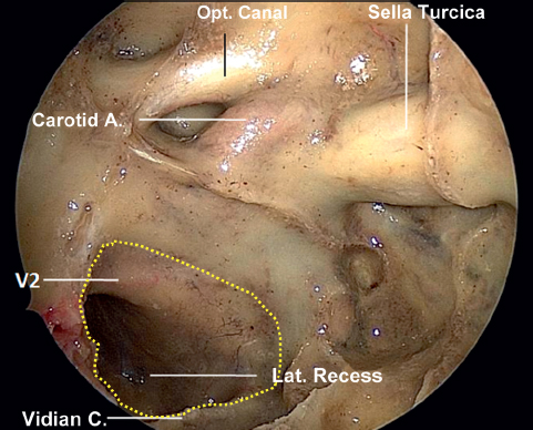
Fig. 37.1 Right sphenoid sinus lateral recess. The dotted line marks the entrance to the right lateral recess of the sphenoid sinus, which is bounded by the vidian canal and V2. A.: artery; C.: canal; Lat.: lateral; Opt.: optical.
 Patient Selection
Indications for an endoscopic transpterygoid approach include lesions located in the lateral sphenoid recess, pterygopalatine fossa, and infratemporal skull base (base of the middle cranial fossa). It is important to identify the extension of the disease preoperatively by means of physical examination, contrast-enhanced high-resolution computed tomography (CT) and magnetic resonance imaging (MRI).6 When treating patients with CSF leaks, it is important to differentiate CSF leak from other sources of rhinorrhea, locating the specific site of the leak and ruling out the presence of increased intracranial pressure. β2-transferrin and to even a greater extent β-trace protein are reliable chemical markers for the presence of CSF.5
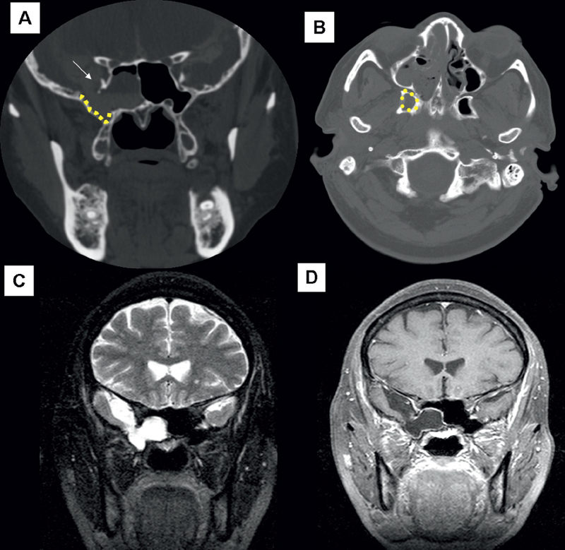
Fig. 37.2 Imaging studies of the lateral sphenoid recess. (A) Coronal high-resolution computed tomography (CT) showing a bony defect indicated by the arrowhead at the lateral recess of the sphenoid sinus (yellow dotted line). (B) Axial high-resolution CT showing the lateral recess of the sphenoid sinus (yellow dotted line). The opposite lateral recess of the sphenoid sinus is normal. (C) T2-weighted magnetic resonance imaging (MRI) showing hyperintense signal in the lateral recess of the sphenoid sinus compatible with cerebrospinal fluid (CSF). (D) T1-weighted MRI with fat suppression showing a low-density lesion in the lateral recess of the sphenoid sinus.
The endoscopic transpterygoid approach addresses lesions that extend to the pterygomaxillary fissure and medial aspect of the middle cranial fossa and infratemporal fossa. An extended lateral reach is possible with the use of an endoscopic Denker’s procedure. This procedure includes an endoscopic medial maxillectomy and the removal of the inferior pyriform aperture and medial maxillary buttress. It is important to have a clear understanding regarding the extension of the lesion (Fig. 37.4).
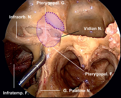
Fig. 37.3 Pterygopalatine fossa contents and location of the lateral recess of the sphenoid. Anatomical dissection shows the contents of the pterygopalatine fossa. The thinner dotted line shows the location of the lateral recess of the sphenoid sinus. The thicker dotted line shows the entrance to the lateral recess of the sphenoid sinus. F.: fossa; G.: ganglion, great; Infraorb.: infraorbital; N.: nerve; Pterygopal.: pterygopalatine.
Related posts:
 Anesthesia in Endoscopic Skull Base and Brain Surgery
The Endonasal Transplanum-Transtuberculum Endoscopic Approach in Pituitary Adenomas
Endoscopic Transnasal Craniectomy and the Resection of Extensive Craniopharyngiomas
External Versus Endoscopic Approaches for Skull Base Malignancies
Pterygopalatine and Infratemporal Fossae
Anatomy of the Orbit and Related Structures
Anesthesia in Endoscopic Skull Base and Brain Surgery
The Endonasal Transplanum-Transtuberculum Endoscopic Approach in Pituitary Adenomas
Endoscopic Transnasal Craniectomy and the Resection of Extensive Craniopharyngiomas
External Versus Endoscopic Approaches for Skull Base Malignancies
Pterygopalatine and Infratemporal Fossae
Anatomy of the Orbit and Related Structures


