CHAPTER 257 Image-Guided Robotic Radiosurgery
The CyberKnife
The CyberKnife is the combination of a real-time image guidance system and lightweight linear accelerator (LINAC) mounted on a highly maneuverable industrial robot. The cascade of properties that emerge from this combination includes the ability to correct in real time, with submillimeter accuracy,1–5 for changes in target position; to treat non-isocentrically, as well as isocentrically; to easily fractionate the treatments; to treat intracranial as well as extracranial targets; and to treat moving targets while preserving a tight dosimetry around the lesion.
The heart of the CyberKnife is its image guidance system. During patient setup and repeatedly during treatment, an x-ray imaging system using amorphous silicon detectors positioned on either side of the patient acquires real-time digital radiographs of the region of interest (Fig. 257-1). The images are automatically registered to a library of digitally reconstructed radiographs (DRRs) derived from the treatment planning computed tomography data set. DRR libraries that anticipate the range of patient orientations likely to be experienced are generated. Transformations and rotations can be estimated on both of the x-ray images based on comparison with the DRRs. The resulting calculations are used to ensure proper initial patient setup and to adjust the aim of the treatment beam in response to small patient movements during treatment sessions. In intracranial applications, the targets are detected and tracked on the basis of information from nearby skull anatomy.5 In the spine, target tracking was initially accomplished with the help of radiopaque fiducials implanted in nearby vertebrae.6 However, recent advances (Xsight Spine Tracking System, Accuray, Inc.) have tracked the nearby vertebral anatomy, thereby alleviating the need for fiducials while maintaining submillimeter lesion tracking.2,3
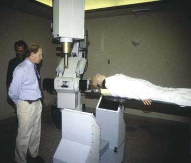
FIGURE 257-1 Photo of the first patient treatment being performed at Stanford in 1994 on the original “alpha” CyberKnife system.
The CyberKnife treatment planning system exploits the robot’s six-degree-of-freedom maneuverability and allows an array of overlapping beams to be superimposed without an isocenter.7 An inverse planning procedure optimizes the set of beam directions and dose to be used on lesions of arbitrary shape and has been demonstrated to deliver dose distributions that closely conform to even highly irregular volumes (see an example of CyberKnife treatment planning in Fig. 257-2).8,9 Even for quite irregularly shaped lesions, this non-isocentric planning can achieve excellent dose homogeneity.8 Similar dose homogeneity may be difficult to achieve with multiple-isocenter methods.9

Perhaps the most important benefit of having successfully eliminated the necessity of the stereotactic frame is the ability to conveniently treat patients in multiple sessions, or fractions. This is a capability that is difficult to match with frame-based radiosurgery. Fractionation is critical for the successful application of radiosurgery outside the central nervous system (CNS) and also has important applications for the treatment of brain and spine lesions that are very near eloquent brain structures and the spinal cord; in such cases, limited fractionation may afford protection to these delicate structures while delivering doses of radiation that control these lesions.10,11 Recently, the American Association of Neurological Surgeons recognized the therapeutic potential of limited fractionation by defining radiosurgery as an ablative procedure that can be performed in one to five fractions.12
Although applications of radiosurgery outside the CNS are of less importance to neurosurgery, the advances of the CyberKnife have expanded radiosurgery out of the confines of neurosurgery, CNS radiation oncology, and neuro-oncology into the general field of oncology. Our most common cancers, prostate and lung malignancies,13,14 can now be targeted with noninvasive fractionated radiosurgery. With prostate tumors, for example, hypofractioned treatment plans allow the delivery of radiation therapy to prostate tumors that is noninvasive (unlike seeds) and has a shorter course (usually five or fewer fractions) than needed with traditional external beam radiation.14,15 The unparalleled ability to track and treat lesions that move with respiration, such as those in the lung and liver, greatly enhances the general utility of the CyberKnife and radiosurgery as a tool in oncology. We believe that it is a neurosurgeon’s role to introduce to our thoracic surgery, surgical oncology, head and neck surgery, and urology colleagues the benefits of radiosurgery and educate them about this tool. Few if any will have had formal exposure to radiosurgery during their training or subsequently.
Clinical Applications of Robotic Image-Guided Radiosurgery
Intracranial Lesions
Frameless radiosurgery has introduced the possibility of treating brain targets with the widest array of penetration trajectories. Rigid frames may exclude the skull base from treatment because the beams cannot pass below the plane of the frame. The CyberKnife can exploit beams penetrating through the splanchnocranium (portion of the skull arising from the first three branchial arches and forming the supporting structure of the jaw) while sparing the brain altogether on the way to extra-axial targets such as vestibular schwannomas or the trigeminal nerve. The ability to hypofractionate treatments may permit safer irradiation of tumors and lesions located close to exquisitely radiosensitive structures such as the optic pathways and brainstem.10,11
The CyberKnife has been used successfully to treat traditional radiosurgery targets, including brain metastases,1 acoustic neuromas,11 arteriovenous malformations,16 and trigeminal neuralgia.17 Outcomes with this frameless radiosurgery approach have been similar to those reported with traditional frame-based treatment. In one study, patients with multiple brain metastases (53 patients, 132 lesions) underwent CyberKnife radiosurgery.18 Overall, 91% of the tumors were controlled with a low rate of radiation necrosis. These findings confirm that frameless CyberKnife radiosurgery can provide control of brain metastases.
The most obvious use of fractionation is around the optic apparatus, such as for meningioma or pituitary adenoma (less commonly metastasis), where doses up to the maximal tolerance of the optic nerves or chiasm rather than the ideal treatment dose for the tumor are frequently delivered with stereotactic radiosurgery. Multiple studies have reported on the safe application of stereotactic radiosurgery for lesions located immediately adjacent to the optic apparatus.10,19,20 In one series of 49 patients with tumors located within 2 mm of the optic pathways, follow-up at a mean of 49 months showed that vision was stable in 38 patients (78%), improved in 8 (16%), and worsened in 3 (6%).10 In the case of visual decline, tumor progression and eventual patient death from the tumor were noted. The treatments were delivered in two to five fractions. These findings suggest that fractionated CyberKnife radiosurgery can be performed safely despite proximity to the optic apparatus and can result in a high rate of tumor control (>92%) at  years’ follow-up.


Stay updated, free articles. Join our Telegram channel

Full access? Get Clinical Tree


