Fever
Acute or subacute onset
Headache
Focal or diffuse symptoms and signs depend on location of infections (see Table 13.2)
Elevated white blood count and erythrocyte sedimentation rate
Increased frequency in immunosuppressed individuals
There are three major sites where infections occur in the CNS: diffusely in the meninges (meningitis), diffusely in the brain (encephalitis), and focally in the brain (abscess) (Fig. 13.1). Table 13.2 gives the major signs and symptoms for infections at these sites. Although there are many different infectious organisms that can infect the meninges and brain, this chapter discusses the most common CNS infections. We will also consider an infection of the peripheral nervous system and a rare infection that breaks conventional rules for infectious diseases.
Table 13.2
Clinical features of major CNS infections
Meningitis | Brain abscess | Encephalitis | |
|---|---|---|---|
Common | Fever | Headache | Fever |
Headache | Mental status changes: confusion, stupor, coma | Headaches, nausea, vomiting | |
Stiff neck | Seizures: generalized or focal | Mental status changes: confusion, stupor, or coma | |
Confusion | 6th nerve palsy | Seizures: generalized or focal | |
Hemiparesis | Hyperreflexia, Babinski signs, or spasticity | ||
Papilledema | Mild stiff neck | ||
Less common | Seizures | Stiff neck | Tremors, choreathetosis-Papilledema |
Stupor or coma | |||
Papilledema |
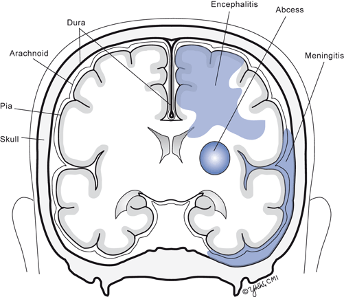
Fig. 13.1
There are three major sites where infections occur in the CNS: diffusely in the meninges (meningitis), diffusely in the brain (encephalitis), and focally in the brain (abscess)
Meningitis
Introduction
Meningitis is due to inflammation of the meninges and is the most common CNS infection. Most infections are due to viruses or bacteria but meningitis also can be caused by fungi, parasites, chemicals, and neoplasms. Viral meningitis is the most common infection of the central nervous system and occurs worldwide with the highest incidence in children and young adults. Viral meningitis is the main cause of a broader term, aseptic meningitis, which refers to meningitis with no laboratory evidence of a bacterial or fungal infection. Viral meningitis occurs mainly in the spring and summer while bacterial meningitis occurs year around. In developed countries, bacterial meningitis occurs equally in children and adults, but is more common in individuals who are immunosuppressed.
Pathophysiology
Enteroviruses, herpes simplex virus type 2, and mumps virus (in countries that do not routinely administer mumps vaccine to children) are the most common etiologies of viral meningitis. Aerobic bacteria, both Gram positive and Gram negative, are the major causes of acute bacterial meningitis. Other bacteria such as Borrelia burgdorferi (Lyme meningitis), Mycobacterium tuberculosis, and Treponema pallidum (neurosyphilis) commonly cause chronic meningitis. The usual route of entry for bacteria and mumps is via the upper respiratory tract where they establish an early asymptomatic infection. Enteroviruses route of entry is oral. Herpes simplex type 2 virus invades mucosa following sexual contact. Most invasive bacterial and viral strains cross though the respiratory or GI epithelial barriers to reach capillaries, veins, and lymphatic channels and then enter the blood. In the blood stream, bacterial characteristics such as large mucopolysaccharide coats hide the bacteria from the reticuloendothelial system allowing them to persist and replicate to high titers, both risk factors for meningitis. Increasing evidence finds that bacteria that cause meningitis can attach to cerebral endothelial cells via specific receptors for each type of bacteria and travel across the barrier inside the endothelial cell to be released in the subarachnoid space. Once within the CSF, the bacteria again replicate and release endotoxin (Gram-negative bacteria) or teichoic acid (Gram-positive bacteria) from their cell walls. Viruses and these molecules stimulate resident macrophages and microglia to release cytokines (especially interleukin-1 and tumor necrosis factor) that in turn recruit neutrophils and mononuclear cells into the CSF from the blood. In bacterial meningitis and most viral meningitis , the organism is confined in the meninges and does not invade the brain parenchyma.
As the inflammation increases in bacterial meningitis but not viral meningitis, the brain becomes irritated and damaged. Endotoxin released from the cell walls of dying bacteria and molecules released from inflammatory cells (such as tumor necrosis factor, neutrophil granule molecules) can pass through the pial lining of the brain, which lacks a blood–brain barrier to invade and kill neurons located at the surface of the cerebral cortex and cerebellum . In addition, the meningeal inflammation can cause vasospasm or thrombosis of arteries and veins passing in the meninges to reach the brain. Occlusion of these vessels leads to cerebral infarctions of the corresponding vascular territory. Thus, while bacteria do not invade the brain, severe brain damage can result from intense meningitis.
In viral meningitis , the host immune system eliminates the virus from the meninges. However, in bacterial meningitis , the immune system does not kill the bacteria, allowing continued bacterial replication until the individual dies unless appropriate antibiotics are administered.
Major Clinical Features
The clinical hallmark of any early meningitis is fever, headache, and stiff neck, and a relatively preserved mental status (Table 13.2). These symptoms have an abrupt onset over a few hours. The headache comes from inflammation of pain fibers along the base of the brain and second and third spinal nerves. The fever may be due to direct activation of the hypothalamus or CSF interleukin-1 released into the CSF by the inflammatory cells. However, all three symptoms are present in only two-thirds of meningitis patients. Patients with bacterial meningitis seldom present with focal neurologic signs but hemiparesis, aphasia , ataxia, and visual loss may develop later in the clinical course. Papilledema is rarely present at onset. Patients with bacterial meningitis can progress from early symptoms to evidence of brain damage within one to a few days.
Major Laboratory Findings
The definite diagnosis of meningitis and establishing whether the meningitis is viral or bacterial is made from the analysis of CSF. When meningitis is suspected, the lumbar puncture (LP) becomes an emergency procedure. Table 13.3 demonstrates the CSF findings in bacterial meningitis and distinguishes them from other CNS infections. Figure 13.2 illustrates the common bacteria that cause meningitis in the USA. In countries that do not give children the Haemophilus influenzae vaccine, H. influenzae meningitis is the most common type for children less than 5 years old. The administration of pneumococcal vaccine is also reducing the incidence of pneumococcal meningitis in children.
Table 13.3
CSF findings in major CNS infectionsa
Opening pressure | White blood cells/mm3 | Predominate WBC type | Protein (mg/dl) | Glucose (mg/dl) | Bacterial or fungal culture | |
|---|---|---|---|---|---|---|
Meningitis | ||||||
Viral | N | 20 − 1000 | Mononuclear | Sl ↑ | Normal | Negative |
Bacterial | N or ↑ | 50 − 5000 | Neutrophils | ↑ | Low | Bacteria |
TB or fungal | ↑ | 50 − 10,000 | Neutrophils and lymphs | ↑ | Low | Sometimes positive |
CNS syphilis | N | 10 − 1000 | Lymphs | ↑ | Normal | Negative |
Brain abscess | ↑ | 0–20 | Lymphs | Normal | Normal | Negative |
Viral encephalitis | Sl ↑ | 10–200 | Lymphs | Sl ↑ | Normal | Negative |
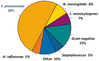
Fig. 13.2
Common bacteria that cause meningitis in the USA
For suspected viral meningitis , the etiology often can be established rapidly for enteroviruses and herpes simplex viruses by polymerase chain reaction (PCR) assay for viral nucleic acid in CSF. Viral culture of CSF obtained early in the meningitis may isolate the virus. Gram staining of CSF sediment identifies the bacterial type over 75 % of the time but does not identify the strain of bacteria or its antibiotic sensitivity pattern. Aerobic bacterial culture of CSF sediment usually isolates the organism and allows subsequent studies to determine antibiotic sensitivity.
Almost all patients with bacterial and many with viral meningitis have an elevated blood WBC count. In bacterial meningitis , neutrophils are elevated and there are elevated numbers of immature cells or a “shift to the left.” The blood erythrocyte sedimentation rate (ESR) and serum C-reactive protein are also elevated.
Neuroimaging usually does not diagnose meningitis. Cranial computed tomography (CT) scans might be indicated before the LP if intracranial masses or acute hydrocephalus is suspected (see section on CSF in Chap. 3 on common neurologic tests). Enhancement of the meninges, especially in the basal cistern area, is commonly seen with gadolinium-enhanced magnetic resonance imaging (MRI) in bacterial meningitis but not viral meningitis. If neurologic complications develop in the patient, neuroimaging may demonstrate communicating or obstructive hydrocephalus, brain infarctions, or focal areas of brain necrosis across the cortical surface.
Principles of Management and Prognosis
For viral meningitis , management is symptomatic with antinausea and analgesic medications. Acyclovir is a drug that prevents herpes simplex virus replication and may shorten the duration of herpes simplex meningitis. Over 95 % of patients with viral meningitis make a full recovery within 1–2 weeks but patients with herpes simplex meningitis may experience relapses over the next 10 years.
For bacterial meningitis , the key to etiologic treatment is the prompt administration of appropriate antibiotics. Without antibiotics, over 95 % of patients die. General principles involved in the use of antibiotics are: (1) the antibiotic should be given as early in the clinical course as possible; (2) the bacteria must be sensitive to the antibiotic administered; and (3) the antibiotic must cross the blood–CSF barrier and achieve sufficient concentration to kill the bacteria. Once the clinical diagnosis of bacterial meningitis is made, immediate treatment with broad spectrum antibiotics begins. Based on the patient’s age, predisposing medical condition, immune status, CSF Gram stain or bacterial antigen tests, and knowledge of types of drug-resistant bacteria in the community, antibiotics are chosen that are likely to kill the CSF bacteria. When the bacterium is grown in the laboratory and antibacterial susceptibilities are determined, the antibiotic regiment can be appropriately modified.
The optimal initial antibiotics to be given constantly changes and one should consult the latest antibiotic recommendations. Currently, many patients with community-acquired meningitis are initially treated with third- or fourth-generation cephalosporins and vancomycin since the incidence of Streptococcus pneumoniae resistance to third-generation cephalosporins is now > 15 % in most communities. In general, the antibiotics should be administered intravenously for 10–14 days. Early administration of corticosteroids for 2–4 days has been shown to reduce the death rate and long-term neurologic sequelae in children and adults with community-acquired bacterial meningitis .
Symptomatic treatment of seizures includes the administration of phenytoin until the patient is discharged. If severe obstructive hydrocephalus develops, a ventriculoperitoneal shunt is required.
Meningitis from Neisseria meningitidis and H. influenzae require chemoprophylaxis of immediate family members and close contacts, who have not received the meningococcal vaccine, with rifampin or ciprofloxacin (adults only) as they are at increased risk of developing meningitis.
Mortality ranges from 5 to 25 % depending on the infecting bacterium, age of patient, and predisposing illnesses. In surviving children, 15 % have language disorders, 10 % mental retardation, 10 % hearing loss, 5 % weakness or spasticity, and 3 % epilepsy. Adults have a similar pattern of neurologic sequelae.
Brain Abscess
Introduction
A brain abscess is a localized intracerebral infection that begins as a focal area of cerebritis and develops into a collection of pus surrounded by a capsule. If the abscess develops outside the brain and around the dura, it is called either a subdural empyema or epidural abscess. Brain abscesses most commonly occur from a bacterial infection but fungi, Mycobacterium tuberculosis, and protozoa can also cause a focal brain infection. Brain abscesses are uncommon with an incidence of about 1 case/100,000 persons per year and develop at all ages but are more common in males.
Pathophysiology
The brain has no normal flora of bacteria or fungi. Microorganisms that cause an abscess reach the brain primarily from adjacent infected sinuses or mastoid air cells, from blood infections, or following head trauma. Common sources of bloodstream infection include infections of lungs (bronchiectasis, empyema, and lung abscess), gastrointestinal tract, urinary system, mouth (dental abscess), heart (acute bacterial endocarditis and cyanotic congenital heart disease), and intravenous drug abuse. In about 20 % of patients, no initial source for the brain abscess can be identified.
The location of the abscess depends on the source. Brain abscesses from frontal sinusitis occur in the frontal lobe adjacent to the infected sinus. Abscesses from mastoiditis develop in the temporal lobe (from upward extension) or occasionally in the cerebellum (from medial extension). The locations of abscesses from a hematogenous route are generally distributed proportional to cerebral blood flow. About three-quarters of brain abscesses are solitary .
The brain abscess begins as a small area of brain infection (cerebritis) often located at the gray–white matter junction of the cerebral cortex. Growth of the organism soon results in expansion of the cerebritis with increasing numbers of neutrophils and mononuclear cells entering the infected site. Necrosis with liquefaction of the center of the abscess then occurs. A variable amount of surrounding cerebral edema contributes to the mass of the abscess. A fibrotic and gliotic response surrounds the abscess forming a capsule but the capsule wall is inadequate to control medial expansion of the abscess. If untreated, the abscess expansion continues until the mass is large enough to cause transtentorial herniation (cerebral hemisphere abscess), foramen magnum herniation (cerebellar abscess), or rupture of the abscess contents into the ventricles (ventriculitis).
Related posts:


Stay updated, free articles. Join our Telegram channel

Full access? Get Clinical Tree


