Intervertebral Disc, T1 Hypointense
Jeffrey S. Ross, MD
DIFFERENTIAL DIAGNOSIS
Common
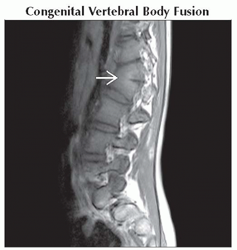
Congenital Vertebral Body Fusion
Degeneration/Calcification
Degeneration/Vacuum Phenomenon
Osteophyte
Instrumentation/Implants
Less Common
Kümmell Disease
Pseudoarthrosis
Neurogenic (Charcot) Arthropathy
Post-Traumatic Instability
Post-Operative Spinal Complications
ESSENTIAL INFORMATION
Helpful Clues for Common Diagnoses
Congenital Vertebral Body Fusion
Linear small low signal disc with “wasp waisting” of vertebral bodies
Degenerative Disc Disease
Calcification or vacuum phenomenon within intervertebral disc
Loss of disc space height, vacuum phenomenon seen as low signal within disc on T1WI
Decreased signal of intervertebral disc on T2WI, loss of central nucleus high signal classic findings of disc degeneration
Osteophyte
Variable appearance due to type of marrow, degree of cortical bone
May show linear low signal on all pulse sequences with dense cortical bone
Larger osteophytes may contain fatty marrow with ↑ T1 signal
Claw-like appearance contiguous with endplates/disc
Instrumentation/Implants
Low signal with distortion and spatial mismapping of signal ⇒ high signal surrounding halo
Small amount of metal artifact may give significant artifact, not visible on CT/plain films
Typical following cervical discectomy
Helpful Clues for Less Common Diagnoses
Kümmell Disease
Nonunited vertebral body fracture undergoes secondary necrosis and collapse
Nitrogen accumulates in fracture cleft
Usually elderly, osteoporotic patients
Low signal on T1WI from gas, variable high signal T2WI, STIR from body
Neurogenic (Charcot) Arthropathy
Destructive arthropathy when pain and proprioception are diminished/lost, while joint mobility is maintained
Preserved bone density, bony debris best seen on CT, helps distinguish from infection
Lumbar spine, rapidly progressive, vacuum phenomenon
Image Gallery
 Sagittal T2WI MR shows L1-2 congenital fusion with rudimentary linear low signal intervertebral disc
 and mild kyphotic angular deformity. and mild kyphotic angular deformity.Stay updated, free articles. Join our Telegram channel
Full access? Get Clinical Tree
 Get Clinical Tree app for offline access

|