Intramedullary Lesion, No Enhancement
Lubdha M. Shah, MD
DIFFERENTIAL DIAGNOSIS
Common
Syringomyelia
Multiple Sclerosis, Spinal Cord
Contusion-Hematoma, Spinal Cord
Acute Transverse Myelitis, Idiopathic
Less Common
Astrocytoma, Spinal Cord
Ependymoma, Cellular, Spinal Cord
Infarction, Spinal Cord
ADEM, Spinal Cord
Cavernous Malformation, Spinal Cord
Rare but Important
Neurenteric Cyst
ESSENTIAL INFORMATION
Key Differential Diagnosis Issues
Imaging of ADEM may be similar to fulminant multiple sclerosis; however, the former is monophasic
Helpful Clues for Common Diagnoses
Syringomyelia
Cystic intramedullary lesions that may be loculated with septations
Enhancement suggests inflammatory or neoplastic lesion
Multiple Sclerosis, Spinal Cord
Enhancement during acute/subacute phase & lasts 1-2 months
Does not reflect disease progression
Cord atrophy usually in late stage & correlates with clinical disability
Contusion-Hematoma, Spinal Cord
Acute: Iso-/hypointense with cord swelling
Chronic: Focal/segmental atrophy
Acute Transverse Myelitis, Idiopathic
Centrally located lesion, 3-4 segments in length
Variable enhancement depending on age
Helpful Clues for Less Common Diagnoses
Astrocytoma, Spinal Cord
10% of cord tumors may show no enhancement
Typically low grade astrocytomas (WHO grade I, II)
Fusiform cord expansion, T2 hyperintensity
Cysts uncommon in nonenhancing tumor subclass
Infarction, Spinal Cord
Early stage may have no T1 signal abnormality
± Patchy enhancement in subacute phase
Cavernous Malformation, Spinal Cord
Absent or minimal enhancement
Helpful Clues for Rare Diagnoses
Neurenteric Cyst
Fluid intensity cystic lesion, typically intradural/extramedullary
Segmentation & fusion anomalies
Image Gallery
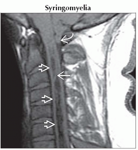 Sagittal T1WI MR shows an elongated, cystic intramedullary lesion
 with CSF signal. This cavity is loculated with septations with CSF signal. This cavity is loculated with septations  . Note the cerebellar tonsillar ectopia . Note the cerebellar tonsillar ectopia  . .Related posts:Stay updated, free articles. Join our Telegram channel
Full access? Get Clinical Tree
 Get Clinical Tree app for offline access

|