3 Lifespan changes in the nervous system
At the end of this chapter you should be able to:
• identify the key changes and time points in the process of neurulation
• explain what is meant by the term ‘cell differentiation’ and what is thought to influence this process in the early stages of neurodevelopment
• describe the cause of the condition known as spina bifida and outline the defects found in the three types of spina bifida: occulta, meningocoele and myelomenigocoele
• discuss the basic ideas behind some of the general theories of ageing
• highlight the main physiological, biochemical and pathological changes associated with ageing.
Neurodevelopment
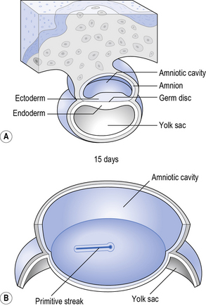
Approximately 12 hours after the moment of fertilisation of the female reproductive tract the zygote divides in two, followed by further divisions every 12 hours or so into four cells, then eight, etc. This process of division continues with little increase in size, until about day 7 when implantation of the embryo occurs. By embryonic day 15 (E15) the structures necessary to sustain the embryo are in place, i.e. the placenta, amniotic cavity and yolk sac (see Fig. 3.1).
Neurulation
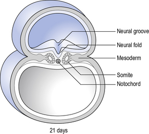
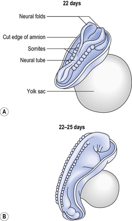
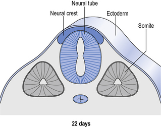
Around day 22 the process of neurulation begins roughly halfway along the neural plate and in the early stages of the process the cranial and caudal ends stay open. The process then spreads along the groove with the cranial neuropore closing around day 25 and the closure of the caudal neuropore taking place around day 27. The closure in both directions occurs in a segmented way and this segmental arrangement is retained in the spinal cord (see Figs 3.2, 3.3 and 3.4).
Embryology of the spinal cord
and these plates are separated by a shallow groove known as the sulcans limitans.
By week 10 the lumen of the neural tube starts to form a small central canal. It is at this point that the alar plate cells develop into the ascending projection neurones and interneurones that will subsequently form sensory pathways and reflex circuits. Simultaneously, the basal plate cells differentiate into the motor neurones and interneurones that will transmit information out of the spinal cord down to the muscles (i.e. the descending projection neurones). The cells in the thoracic segment develop into the sympathetic preganglionic neurones, while the cells in the sacral segment develop into the parasympathetic preganglionic neurones – thereby forming both parts of the autonomic nervous system (see Chapter 1).


