Chapter 30 Lower Back and Lower Limb Pain
Lower back pain is one of the most common reasons for neurological and neurosurgical consultation. The cost to society is huge, with estimates of up to $80 billion per year in direct and indirect healthcare costs and loss of productivity. In Switzerland, low back pain consumes 6.1% of the total healthcare budget and up to 2.3% of their GDP (Wieser et al., 2010). In many of the patients who present with lower back pain, the pain either developed or was exacerbated as a result of occupational activity. Lower limb pain is a common accompaniment to lower back pain but can occur independently.
Related Anatomy and Physiology
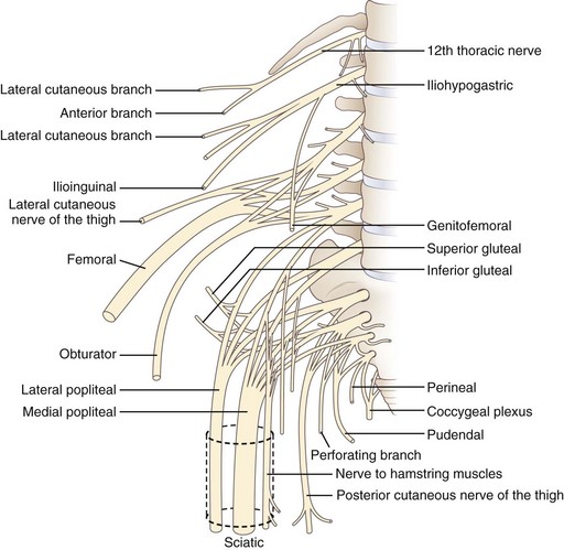
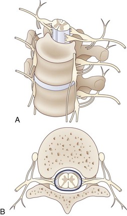
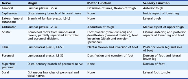
The lumbosacral spinal cord terminates in the conus medullaris at the level of the body of the L1 vertebra (Fig. 30.1). The motor and sensory nerve roots from the lumbosacral cord form the cauda equina. From there, the motor and sensory nerve roots unite at the dorsal root ganglion to form the individual spinal nerves. These anastomose in the lumbosacral plexus (Fig. 30.2), from which run the major nerves supplying the leg (Table 30.1).

Fig. 30.2 Anatomy of the lumbosacral plexus.
(Reprinted with permission from Bradley, W.G., 1974. Disorders of the Peripheral Nerves. Blackwell, Oxford, p. 29.)
Causes of lower back pain without leg pain include:
Causes of lower back plus lower limb pain include:
Important causes of leg pain without low back pain include:
Diagnosis
History and Examination
The history should focus first on features of the back and leg pain:
 Associated motor and sensory symptoms
 Exacerbating and remitting factors
 History of predisposing factors (e.g., trauma, cancer, osteoporosis)
Differential Diagnosis of Lower Back and Leg Pain
The differential diagnosis of lower back and leg pain can be addressed as shown in Tables 30.2 through 30.5. Classification into mechanical and neuropathic categories is useful to narrow the scope of diagnostic considerations. The possibility of non-neurological causes should always be kept in mind.
Table 30.2 Classification of Lower Back and Lower Limb Pain
| Type | Examples |
|---|---|
| Mechanical pain | |
| Neuropathic pain | |
| Non-neurologic pain |
Table 30.3 Differential Diagnosis of Lower Back and Leg Pain
| Disorder | Clinical Features | Diagnostic Findings |
|---|---|---|
| Radiculopathy | Back pain radiating into leg in a dermatomal distribution. Sensory loss and motor loss are in a root distribution. Increased pain with coughing or straining. | Suspected when neuropathic pain radiates from back down into leg in a single root distribution. Disk or mass can be seen on MRI or CT. Zoster and diabetes can cause radiculopathy without abnormal studies. |
| Plexopathy | Back and leg pain with a neuropathic character, dysesthesias, burning, or electric sensation. Back pain can develop when cause is mass lesion in region of plexus. | Suspected when patient has leg pain in more than one peripheral nerve or root distribution. MRI of plexus or CT of abdomen and pelvis can show mass or hematoma. |
| Spinal stenosis | Pain in lower back, buttocks, and legs, especially with standing, walking, and lumbar spine extension. | MRI or CT shows obliteration of subarachnoid space. |
CT, Computed tomography; MRI, magnetic resonance imaging.
Table 30.4 Differential Diagnosis of Isolated Lower Back Pain
| Disorder | Clinical Features | Diagnostic Findings |
|---|---|---|
| Sacroiliac joint inflammation | Pain lateral to spine where sacrum inserts into top of iliac bone. Pain is exacerbated by movement and pressure but does not radiate down leg. | Clinical diagnosis. Radiographs can show degenerative changes in joint. Bone scan shows increased uptake in region. |
| Facet pain | Unilateral or bilateral paraspinal pain without radiation. Pain is increased by spine motion, especially extension. | Clinical diagnosis. Radiographs can show facet degeneration. |
| Ovarian cyst or cancer | Pain in hip and lower back, often but not always extending into lower quadrant. Bowel disturbance may develop with advanced disease. | Abdominal and pelvic CT shows mass lesion in ovary. |
| Endometriosis | Usually pelvic pain but occasionally pain in back and legs. Pain is often timed to menses. | Diagnosis suspected during pelvic exam. Vaginal ultrasound is supportive. Laparoscopy is diagnostic. |
| Retroperitoneal mass, abdominal aortic aneurysm, abscess, hematoma | Pain in back. May be bilateral to spine. May be associated with superimposed neuropathic pain in cases with plexus or proximal nerve involvement. | CT or MRI shows hematoma, aneurysm, eroding vertebral bodies, or abdominal mass. |
| Urolithiasis | Pain in upper to mid-back laterally that may radiate to groin. No radiation into leg. | Radiographs may show stones. Intravenous pyelography typically shows obstruction of flow. Contrasted abdominal CT usually shows the stone and obstruction. |
| Diskitis | Pain in lower back exacerbated by movement. Some patients may have radiation of pain to abdomen, hip, or leg. | MRI shows characteristic changes in disk and surrounding tissues. |
Table 30.5 Differential Diagnosis of Isolated Leg Pain
| Disorder | Clinical Features | Diagnostic Findings |
|---|---|---|
| Peroneal neuropathy | Loss of sensation on dorsum of foot. Weakness of foot and toe dorsiflexion. | Slowed nerve conduction velocity across region of entrapment, usually at fibular neck. EMG may show denervation in peroneal-innervated muscles, especially tibialis anterior, without involvement of short head of biceps femoris. |
| Femoral neuropathy | Pain and sensory loss in anterior thigh, often with weakness of quadriceps and suppression of knee reflex. | NCS can sometimes be performed but may be technically difficult. EMG may show denervation in a distribution limited to femoral nerve. |
| Piriformis syndrome | Pain from back or buttock down posterior thigh. Pain is exacerbated by sitting or climbing stairs. Stretch of piriformis (flexion and adduction of the hip) worsens pain. | Clinical diagnosis. Pain radiating down leg in a sciatic nerve distribution. Exacerbation of pain by flexion and adduction of hip. EMG and NCS may show proximal sciatic nerve damage. |
| Meralgia paresthetica (lateral femoral cutaneous nerve dysfunction) | Pain and loss of sensation of lateral femoral cutaneous nerve on lateral aspect of thigh. | Clinical diagnosis. NCS is difficult to perform on this nerve. |
| Claudication | Pain in thigh and lower leg with exertion. Pain does not occur with lumbar spine extension. | Suspected with exertional leg pain without back pain. Ultrasonography or angiography confirms arterial insufficiency. |
| Plexopathy | Back and leg pain that has a neuropathic character. Dysesthesias, burning, or electric sensation. Plexitis has no associated back pain. | Suspected when a patient has leg pain in more than one peripheral nerve distribution. MRI of plexus or CT of abdomen can show a structural lesion in some patients. |
| Radiculopathy | Pain largely in one dermatomal distribution. May be motor and reflex loss. Most patients have back pain, but not all. | Suspected with pain radiating down one leg with or without back pain. Best imaged by MRI or postmyelographic CT. |
CT, Computed tomography; EMG, electromyography; MRI, magnetic resonance imaging; NCS, nerve conduction studies.
Some basic guidelines for the differential diagnosis of lower back and leg pain are as follows:
 Pain confined to the lower back generally is caused by a low back disorder.
 Pain confined to the leg usually is caused by a leg disorder, although neuropathic pain from lumbar spine disease can radiate down the leg without back pain in a minority of patients.
 Pain in both the low back and the leg usually is caused by lumbar radiculopathy or, less commonly, lumbosacral plexopathy.
 Clinical abnormalities confined to one nerve root distribution usually are caused by intervertebral disk disease or lumbosacral spondylosis producing radiculopathy.
 Clinical abnormalities that involve several nerve distributions usually are caused by plexus lesions, with cauda equina lesions being the alternative diagnosis.
 Bilateral lesions suggest proximal damage in the spinal canal affecting the roots of the cauda equina.
 Impairment of bladder control indicates either a cauda equina lesion or, less commonly, a bilateral sacral plexopathy.
 Presence of more than one lesion may complicate neurological localization.
 Non-neurological causes of lower back pain are possible and particularly include urolithiasis, abdominal aortic aneurysm, and other intraabdominal pathological processes.
 Multiple lesions can make the differential diagnosis more difficult. For example, radiculopathies at two or more levels may look like a plexopathy or peripheral neuropathic process.
Stay updated, free articles. Join our Telegram channel

Full access? Get Clinical Tree


