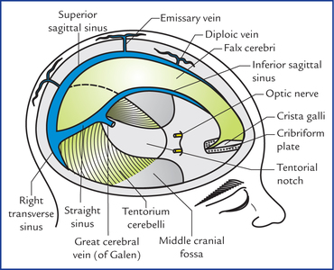
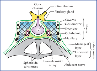
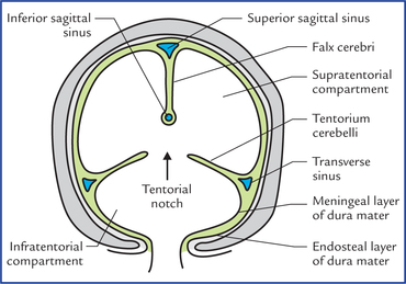
Meninges And Cerebrospinal Fluid
Meninges
The brain and spinal cord are enclosed in three protective membranes called meninges. From without inwards these are: (a) dura mater, (b) arachnoid mater, and (c) pia mater.
The dura mater is mesodermal in origin while arachnoid and pia mater are ectodermal in origin (derived from neural crests).
The dura mater is the thick outermost covering of the brain and spinal cord. The part enclosing the brain is called cranial/cerebral dura, and the part around the spinal cord, the spinal dura. It is very tough opaque inelastic membrane of fibrous tissue (Gk. dura = tough, mater = mother). It is also called pachymeninx (pachy = thick).
The arachnoid mater (Gk. arachnoid = cobweb like, mater = mother) is a delicate avascular membrane deep to dura mater. Many thread-like trabeculae extend from it to the pia mater.
The pia mater (Gk. pia = tender, mater = mother) is a thin transparent vascular membrane closely adherent to the surface of the brain and spinal cord.
The arachnoid mater and pia mater together are termed leptomeninges (Gk. lepto = thin).
The intracranial arrangement of these membranes differs from that in the vertebral canal and are therefore described separately.
The spinal meninges are described in Chapter 7).
Intracranial Meninges
Dura mater
The dura mater in the cranium (cranial dura) consists of two layers: an outer endosteal layer and an inner meningeal layer. These two layers are firmly adherent to each other everywhere except, (a) where they split to enclose the venous sinuses, and (b) where the inner layer is folded to form the dural septa.
The endosteal layer is attached to the inner surfaces of the cranial bones and is continuous through the sutural ligaments, and around the margins of the foramina with the periosteum on the external surface of the cranium.
The meningeal layer is a strong fibrous membrane and becomes continuous with the spinal dura at the foramen magnum. It ensheathes the cranial nerves in their osseous foramina and fuses externally with epineurium; the sheaths of the optic nerves fuse with the ocular sclera.
Dura! septa or folds (Figs 16.1, 16.2)
Fig. 16.2 Schematic coronal section of skull showing division of cranial cavity into three compartments by falx cerebri and tentorium cerebelli. Each half of the supratentorial compartment contains the cerebral hemisphere, whereas the infratentorial compartment contains the cerebellum and brainstem.
The four important dural septa are:
Falx Cerebri
Falx cerebri is a large sickle-shaped vertical fold of dura mater which dips into the longitudinal fissure, between the two cerebral hemispheres.
Its narrow anterior end is attached to the crista galli and its broad posterior end to the upper surface of the tentorium cerebelli. Its convex upper border is attached to the vault of skull along the sagittal suture and its lower border is free and concave downwards.
The falx cerebri encloses three dural venous sinuses, viz.
Tentorium cerebelli
Tentorium cerebelli is tent-like semilunar fold of dura mater which forms the sloping roof of the posterior cranial fossa, between the cerebellum below and the occipital lobes of cerebral hemispheres above. It prevents the cerebellum from being compressed by the heavy cerebrum.
It has two borders: (a) an outer convex attached border, and (b) an inner concave free border. The inner border bounds an oval space, the tentorial notch or the door of tentorium through which passes the midbrain to connect the hindbrain with the forebrain (Fig. 16.2).
The inner free border is ‘U’-shaped and its anterior ends are attached to the anterior clinoid processes.
Tentorium cerebelli contains four dural venous sinuses, two on either side:
Falx cerebelli
Falx cerebelli is a small sickle-shaped fold of dura mater which intervenes between the two cerebellar hemispheres posteriorly. It is attached to the internal occipital crest by its outer border, and encloses the occipital venous sinus.
Arterial supply of dura mater
The dura is supplied by numerous branches of the internal carotid, ascending pharyngeal, maxillary, occipital and vertebral arteries.
N.B. Middle meningeal artery is the largest of the meningeal arteries and from clinical point of view is the most important for it is often damaged in head injuries.
Nerve supply of dura mater
The nerve supply of dura mater is derived mainly from three sources:
• The supratentorial dura is supplied by the meningeal branches from the three divisions of the trigeminal nerve:
1. In the anterior cranial fossa by meningeal branches of the anterior and posterior ethmoidal nerves.
2. In the middle cranial fossa by meningeal branches of maxillary and mandibular nerves.
• The infratentorial dura is supplied by ascending meningeal branches of upper three cervical nerves.
Dural venous sinuses
The dural venous sinuses are formed in following two ways: (a) by separation of the two layers of cerebral dura, and (b) by reduplication of the meningeal layer of dura (Fig. 16.2).
• Have no valves, hence the blood can flow in either direction in the sinuses,
• Are devoid of smooth muscle fibres in their walls,
• Drain finally into the internal jugular veins,
• Have cerebral, diploic and some meningeal veins as their tributaries,
• Communicate via valveless emissary veins with the extracranial veins through skull foramina.
The dural venous sinuses are classified into two types, unpaired and paired (Table 16.1).
Table 16.1
Classification of dural venous sinuses
Unpaired | Paired |
• Superior sagittal sinus | • Sphenoparietal sinuses |
• Inferior sagittal sinus | • Cavernous sinuses |
• Straight sinus | • Superior petrosal sinuses |
• Occipital sinus | • Inferior petrosal sinuses |
• Anterior intercavernous sinus | • Transverse sinuses |
• Posterior intercavernous sinus | • Sigmoid sinuses |
Unpaired sinuses
Superior sagittal sinus lies in the attached border of the falx cerebri and extends anteroposteriorly from the foramen caecum where it communicates with the nasal veins to the internal occipital protuberance, where it usually turns to the right side to continue as the right transverse sinus.
Large clusters of arachnoid villi (arachnoid granulations), concerned with the absorption of CSF project into the venous lacunae of the SSS (the clefts between the two layers of dura mater along side the SSS).
The 8 to 12 superior cerebral veins ascend with slight anterior inclination, and traverse the subdural space to drain into the superior sagittal sinus and its lacunae.
Inferior sagittal sinus lies in the posterior two-third of the lower free border of falx cerebri and receives the veins from the medial surfaces of the cerebral hemispheres. At the junction of falx cerebri with the tentorium cerebelli, it is joined by the great cerebral vein to form the straight sinus.
Straight sinus runs along the line of attachment of falx cerebri with the tentorium cerebelli. At the external occipital protuberance it usually turns to the left to continue as the left transverse sinus.
Occipital sinus is the smallest sinus and situated along the attached margin of the falx cerebelli. It extends downwards from the confluence of the sinuses to the foramen magnum, where it communicates with the internal vertebral venous plexus.
N.B. At the external occipital protuberance the superior sagittal sinus communicates with the left transverse, occipital and straight sinuses to form, what is termed confluence of sinuses.
The shapes of various dural folds, and sinuses enclosed in them are summarized in Table 16.2.
Table 16.2
Shapes of dural folds and enclosed venous sinuses
Fold | Shape | Venous sinuses enclosed |
Falx cerebri | Sickle-shaped | Superior sagittal, inferior sagittal and straight sinuses |
Tentorium cerebelli | Tent-shaped (semilunar) | Transverse and superior petrosal sinuses |
Falx cerebelli | Sickle-shaped | Occipital sinus |
Diaphragma sellae | Horizontal fold | Anterior and posterior intercavernous sinuses |
Paired sinuses
Sphenoparietal sinuses (Fig. 16.4)
These small sinuses lie along the lesser wing of sphenoid and drain into the cavernous sinus.
Cavernous sinuses (Figs 16.3, 16.4)
Cavernous sinuses are situated one on either side of the sella turcica and, the body of sphenoid. Each sinus is a short wide venous channel measuring 2 cm anteroposteriorly and 1 cm transversely. It extends from superior orbital fissure anteriorly to the apex of petrous temporal bone posteriorly. The cavernous sinus is so named because it is traversed by a network of trabeculae which subdivide the cavity into numerous communicating caverns lined by endothelium. The two sinuses communicate with each other through anterior and posterior inter-cavernous sinuses which lie in the anterior and posterior margins of the diaphragma sellae, and pass anterior and posterior to the stalk of the pituitary gland respectively.

Stay updated, free articles. Join our Telegram channel

Full access? Get Clinical Tree

