4 Meninges
Cranial Meninges
Dura mater
The terminology used to describe the cranial dura mater varies among different authors. It seems best to regard it as a single, tough layer of fibrous tissue that is fused with the endosteum (inner periosteum) of the skull, except where it is reflected into the interior of the vault or is stretched across the skull base. Wherever it separates from the periosteum, the intervening space contains venous sinuses (Figure 4.1).
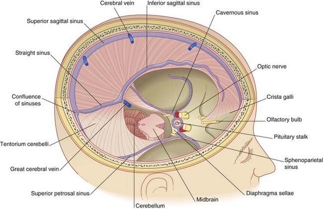
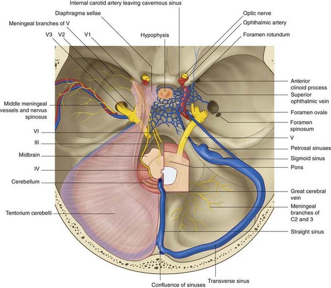
The crescentic tentorium cerebelli arches like a tent above the posterior cranial fossa, being lifted up by the falx cerebri in the midline. The attached margin of the tentorium encloses the transverse sinuses on the inner surface of the occipital bone and the superior petrosal sinuses along the upper border of the petrous temporal bone. The attached margin reaches to the posterior clinoid processes of the sphenoid bone. Most of the blood from the superior sagittal sinus usually empties into the right transverse sinus (Figure 4.2).
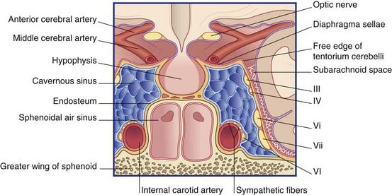
The free margin of the tentorium is U-shaped. The tips of the U are attached to the anterior clinoid processes. Just behind this, the two limbs of the U are linked by a sheet of dura, the diaphragma sellae, which is pierced by the pituitary stalk. Laterally, the dura falls away into the middle cranial fossae from the limbs of the U, creating the cavernous sinus on each side (Figure 4.3). Behind the sphenoid bone, the concavity of the U encloses the midbrain.
Innervation of the cranial dura mater
The dura mater lining the supratentorial compartment of the cranial cavity receives sensory innervation from the trigeminal nerve. That lining the anterior cranial fossa and anterior part of the skull vault is supplied by its ophthalmic branch; that lining the middle cranial fossa and midregion of the vault is mainly supplied by the nervus spinosus (Figure 4.2). This nerve leaves the mandibular nerve outside the foramen ovale, to return via the foramen spinosum and accompany the middle meningeal artery and its branches. Stretching or inflammation of the supratentorial dura gives rise to frontal or parietal headache.
The dura mater lining the infratentorial compartment is supplied by branches of upper cervical spinal nerves entering the foramen magnum (Figure 4.2). Occipital and posterior neck pains accompany disturbance of the infratentorial dura. Acute meningitis involving posterior cranial fossa meninges is associated with neck rigidity and often with head retraction brought about by reflex contraction of the posterior nuchal muscles, which are supplied by cervical nerves. Violent occipital headache follows subarachnoid hemorrhage (Ch. 35), where free blood swirls around the hindbrain.
Meningeal arteries
Embedded in the endosteum of the skull are several meningeal arteries whose main function is to supply the diploë (bone marrow). Much the largest is the middle meningeal artery, which ramifies over the inner surface of the temporal and parietal bones. Tearing of this artery, with its accompanying vein, is the usual source of an extradural hematoma (Clinical Panel 4.1).


