CHAPTER 366 Microsurgery of Paraclinoid Aneurysms
Paraclinoid aneurysms are defined as aneurysms arising from the internal carotid artery (ICA) in close proximity to the anterior clinoid process. Using the broadest definition, such aneurysms could include those arising from the intracavernous, clinoidal, ophthalmic, and posterior communicating ICA segments.1–3 The ensuing chapter, however, addresses only those lesions arising beyond the venous lumen of the cavernous sinus and proximal to the origin of the posterior communicating artery—the clinoidal and ophthalmic segments. More proximal and distal aneurysm variants are discussed in other chapters.
Anatomy
Osseous Anatomy
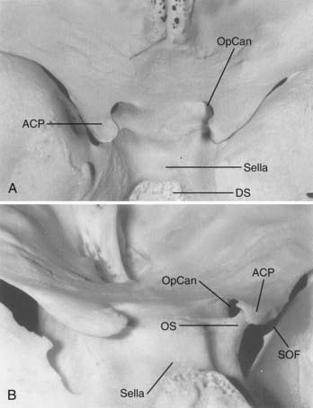
The anterior clinoid process (ACP), formed by the medial extension of the lesser wing of the sphenoid bone, provides a bony roof to the superior orbital fissure (SOF) and the anterior cavernous sinus (Fig. 366-1). The optic strut extends from the inferomedial surface of the ACP to the body of the sphenoid bone, separating the optic canal from the SOF. The ACP and optic strut define and obstruct access to the anterior and lateral borders of the ascending ICA as it exits the cavernous sinus to enter the subarachnoid space.
Dural Anatomy
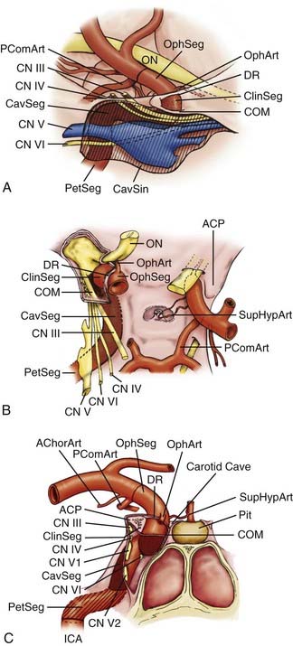
The relevant dural structures in this region represent dural reflections off the ACP, which primarily include the falciform ligament, the (distal) dural ring, and the carotid-oculomotor membrane (COM; Fig. 366-2).4 The dural ring represents the circular point of attachment of the dura to the ICA as the vessel penetrates the dura to enter the subarachnoid space. This dural attachment extends radially from the ICA and is continuous with the periosteum covering the superomedial aspect of the anterior clinoid process, the diaphragma sella, and the floor of the optic canal.5–9 The plane of the dural ring slopes downward from anterior-to-posterior and lateral-to-medial directions. The subarachnoid space diverticulum medial to the ICA created by this slant is called the carotid cave.6–10 The COM is formed by dura from the inferior surface of the ACP that extends from the ICA medially to the oculomotor nerve laterally.4 This membrane marks the point of exit of the ICA from the cavernous sinus main lumen.
Neural Structures
The cranial nerves within the cavernous sinus (CN III, IV, V, and VI) are rarely clinically affected by paraclinoid aneurysms because these lesions generally project superiorly or medially away from the superior orbital fissure and lateral cavernous sinus wall (see Fig. 366-2). Nonetheless, their close proximity to the ACP makes knowledge of their anatomy mandatory so as to avoid injury during its removal.
Vascular Structures
Arterial Segments
The paraclinoid ICA is herein composed of two regions—the clinoidal and ophthalmic segments (see Fig. 366-2).11 The clinoidal segment, the distal portion of the anterior ascending vertical segment of the cavernous carotid artery, is located below and medial to the ACP, above the major venous lumen of the cavernous sinus. This region is a transitional ICA segment found between the COM and the dural ring.4,9,10 The clinoidal segment is located neither within the venous channels of the cavernous sinus nor within the subarachnoid space, and is essentially “interdural.”4,9,10 The ophthalmic segment, the distal portion of the paraclinoid ICA region, lies entirely within the subarachnoid space above and medial to the ACP. Beginning at the dural ring, the ophthalmic segment extends to the origin of the posterior communicating artery, and generally represents the longest subarachnoid ICA segment.11
Arterial Bends and Branches
Saccular aneurysms typically form at points of hemodynamic stress where a bend in the vessel and a branch site coincide.12 Two major bends in the paraclinoid ICA region predispose this region to aneurysm formation. The first bend, seen best on lateral angiogram, is the posteriorly projecting turn that begins at the anterior genu of the cavernous segment and continues as the vessel ascends through the dural ring. This bend creates a strong superior vector on the anterior and dorsal wall of the clinoidal and ophthalmic segments. A less conspicuous second bend, seen best from an anteroposterior or dorsal view, is the gentle lateral-to-medial-to-lateral curve beginning at the anterior genu of the cavernous segment and continuing as the ICA travels toward its terminal bifurcation. This medially projecting hemodynamic vector places significant stress upon the medial aspect of the clinoidal and ophthalmic ICA segments.
The ophthalmic segment harbors several prominent arterial branches that predispose this region to aneurysm formation. The ophthalmic artery is the most prominent and clinically significant branch, and typically arises from the dorsomedial carotid surface just above the dural ring and below the inferolateral portion of the optic nerve.13,14 This branch projects anterolaterally to run through the optic canal beneath the optic nerve, and supplies the optic nerve through perforating branches and the retina through the central retinal artery. The superior hypophyseal artery is the second major branch (or series of branches), usually arising from the medial or inferomedial ICA surface just distal to the dural ring, medial and unrelated to the take-off of the ophthalmic artery along the medial-to-lateral ICA bend within the ophthalmic segment.15,16 This vessel projects medially to supply the superior aspect of the pituitary stalk and gland, a portion of the cavernous sinus dura, and the posterior optic nerve and chiasm.
The clinoidal segment is usually devoid of named arterial perforators. On occasion, however, the ophthalmic or superior hypophyseal arteries originate from this segment, typically reaching their end organs through alternate anatomic pathways. The ophthalmic artery, for example, can originate from the distal cavernous segment or clinoidal segment in up to 10% of cadaveric specimens, usually entering the orbit through the SOF or an orifice within the optic strut.4,17,18
Aneurysm Classification
Clinoidal Segment Aneurysms
Two clinoidal segment aneurysm variants can be differentiated according to their site of origin, direction of projection, and relationships to arterial bends and branches and the adjacent dural and osseous structures within the segment (Fig. 366-3).
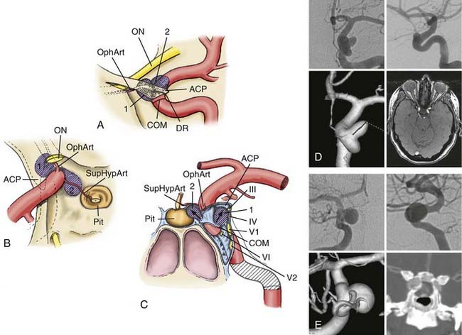
Anterolateral Variant
The anterolateral variant arises from the anterolateral surface of the clinoidal segment as it obliquely ascends toward the dural ring underneath the ACP.1 The superiorly and slightly medially directed course of the segment promotes an aneurysm that expands lateral and anterior to the ascending ICA, superiorly projecting toward and into the ACP. When small, the anterolateral variant may erode the optic strut and undersurface of the ACP to cause monocular visual loss from ipsilateral optic nerve compression within the optic canal. Larger lesions may secondarily compress the visual system (nerve or chiasm) within the subarachnoid space after extension through the dura medial to the ACP. These aneurysms can sometimes arise in association with a clinoidal segment origin of the ophthalmic artery but are more often associated just with the hemodynamic stress associated with the anterior ascending vertical segment of the cavernous ICA.
Medial Variant
The medial variant extends from the medial surface of the clinoidal segment and enlarges toward the sphenoid sinus and sella as the ICA turns from lateral to medial to lateral during its ascent toward and through the dural ring.1 Initially, this aneurysm type expands beneath the diaphragma sella into the pituitary fossa. Gradual enlargement may cause hypopituitarism; rarely, aneurysm rupture into the pituitary gland may simulate pituitary apoplexy. Extension and rupture into the sphenoid sinus may cause life-threatening epistaxis. Large or giant lesions may extend through the diaphragma sella to enter the subarachnoid space. Visual loss from this aneurysm type does not occur with small lesions, but field cuts resembling those of pituitary tumors may occur with large or giant lesions that extend into the suprasellar space.
Ophthalmic Segment Aneurysms
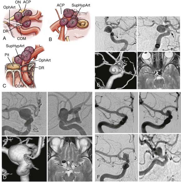
Three ophthalmic segment aneurysm variants can be differentiated according to their site of origin, direction of projection, and relationships to arterial bends and branches and the adjacent dural and osseous structures within the segment (Fig. 366-4).
Ophthalmic Artery Aneurysms
Ophthalmic artery aneurysms arise immediately distal to and in relation to the origin of the ophthalmic artery.1,2,19–22 These lesions typically arise along the posterior bend of the ICA just distal to the ophthalmic artery and dural ring, project dorsally or dorsomedially, and elevate the lateral edge of the nerve against the falciform ligament. The pressure from the falciform ligament against the superior surface of the nerve results in an inferior nasal field defect. A monocular inferior nasal field defect is initially produced; as the lesion enlarges, however, the entire nasal field can become affected, followed by a superior temporal field loss in the contralateral eye. If the clinical course is protracted, ipsilateral blindness with marked contralateral deficits can occur. Larger ophthalmic artery aneurysms tend to thicken or even calcify along their anterior portion, a factor that must be accommodated during clip placement.
Superior Hypophyseal Artery Aneurysms
Superior hypophyseal artery aneurysms arise in association with the superior hypophyseal artery along the medial surface of the ICA, usually just distal to the dural ring.2,20 There are two main variants. The parasellar variant projects inferiorly toward the sella into the carotid cave, whereas the suprasellar variant projects superiorly into the suprasellar space. Parasellar superior hypophysal artery aneurysms are usually asymptomatic. Because they project into the dura of the sella or cavernous sinus, purely parasellar aneurysms are unlikely to rupture.1 Their medial projection and proximity to ICA perforators also makes them more difficult to treat. Suprasellar or large lesions have a much higher tendency to rupture. When large or giant, these aneurysms tend to elevate the optic chiasm and produce visual changes and relationships to the visual system similar to those seen with pituitary tumors. Larger superior hypophyseal artery aneurysms tend to develop thickenings or calcifications along the anterior-medial aspect, near their origin from the ICA, similar to ophthalmic artery aneurysms. Because these lesions extend medially underneath the optic chiasm, they can result in bilateral visual field defects.
Many small lesions burrow inferiorly and medially toward and beneath the diaphragma sella, expanding the carotid cave. These lesions, termed parasellar variant superior hypophyseal artery aneurysms, have also been called carotid cave aneurysms.1,2,7,20 When small, the fundus of this variant is invested by adjacent lateral parasellar dura, and their risk for subarachnoid hemorrhage is quite low. With growth, however, these lesions expand superomedially above the diaphragma sella into the suprasellar space, where the hemorrhage risk becomes greater. A second type, the suprasellar variant superior hypophyseal artery aneurysm, projects medially above a shallow carotid cave and expands into the medial suprasellar space above the diaphragm, without investment by the parasellar dura. Superior hypophyseal artery lesions cause their visual compression by expansion into the suprasellar space and elevation of the optic chiasm, producing superior bitemporal or other patterns of visual loss more suggestive of pituitary tumors.


Stay updated, free articles. Join our Telegram channel

Full access? Get Clinical Tree


