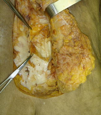
Fig. 45.1
Skin incision for mini-open lumbar fusion and percutaneous pedicle screw
45.2 The Lumbar Muscles and Their Fasciae [2]
The posterior lumbar musculature refers to those muscles that are located behind the plane formed by the transverse processes, and from the surgical point of view, we have three important muscles: the multifidus, the longissimus, and the iliocostalis muscles, with the latter two forming what we know as erector spinae muscle in the lumbar region.
The multifidus muscle is the larger and more central of the posterior lumbar muscles. It consists of a repeated series of fascicles that originate in laminae and spinous processes of the lumbar vertebrae and exhibit a uniform caudal attachment.
The key morphological characteristic of the multifidus is that it has a segmental arrangement. Each lumbar vertebra gives origin to a group of fascicles that radiate from its spinous process and is anchored inferiorly to mammillary processes of the inferior vertebrae, the sacrum, and the iliac crest. From the surgical standpoint, the multifidus muscle occupies a space delimited anteriorly by the lamina, medially by the middle line and laterally by the lateral border of the facets joints.
The lumbar erector spinae muscle lies laterally to the multifidus muscle and shapes the prominent contour of posterolateral lumbar region. It consists of two muscles: the longissimus and iliocostalis.
The longissimus muscle is composed of five fascicles originating in the processes accessories and part of the transverse processes and a caudal iliac spine insertion and is located lateral to the multifidus muscle occupying the spaces previously delimited by transverse apophysis.
The iliocostalis muscle consists of four fascicles that overlap L1 to L4. Rostrally, each fascicle originates from the tip of the transverse processes and the medial part of the thoracolumbar fascia. Integrated into the iliac crest, it has a similar arrangement to the longissimus fascicles with the distinction they are more lateral.
From the surgical point of view, two fasciae are important: the more superficial thoracolumbar fascia and the deeper erector spinae muscle fasciae. The caudal portion of the tendon of the muscle fibers of longissimus and iliocostalis thoracis forms the latter fascia. This aponeurosis does not connect to the lumbar erector spinae muscle fibers (Fig. 45.2).


Fig. 45.2
Thoracolumbar fascia and the deeper erector spinae muscle fasciae
The architecture of the multifidus muscles and longissimus creates a natural cleavage plane around the lateral border of the superior articular facet. For minimally invasive approaches, it is important to identify this avascular plane. In 1988, Wiltse described this cleavage plane as it deviates from the midline in the cranial-caudal direction.
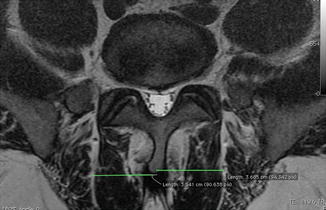
Daniel Palmer et al. [13] detailed that the mean measurements of the cleavage plane between the multifidus and longissimus from the midline significantly differed between all disc levels. At L5–S1, the mean distance was 37.8 mm; at L4–L5, 28.4 mm; at L3–L4, 16.2 mm; at L2–L3, 10.4 mm; and at L1–L2, 7.9 mm. The mean female distances were significantly greater than males (2 mm) on both sides of L5–S1 only (Fig. 45.3).


Fig. 45.3
Measurements of the cleavage plane between the multifidus and longissimus from the midline
45.3 Indications and Contraindications
The indications for mini-open arthrodesis are no different from traditional techniques and patients fall in three main groups:
1.
Primary segmental instability due to spondylolisthesis mainly grades I and II, as a result of disc degenerative disease (discogenic pain) or associated with a herniated disc and/or lumbar canal stenosis
2.
Postdiscectomy due to secondary segmental instability after laminectomy or facetectomy
3.
Patients whose decompressive surgery could lead to extensive iatrogenic instability
The main relative contraindications are:
Spondylolisthesis grades III and IV, three or more levels disc disease requiring surgery, severe osteoporosis, and morbid obesity.
45.4 Preoperative Planning
A thorough anatomical knowledge of all associated structures is key to preoperative planning. The surgeon must know the average distance from the midline to the intermuscular interval between the multifidus muscle and longissimus muscle. Having the ability to tailor the preoperative magnetic resonance examination is important for marking the cutaneous incision surgery by the paramedian intermuscular approaches described by Wiltse and increasingly used for performing minimally invasive fusion.
To achieve surgically the intermuscular plane between the muscle multifidus and longissimus as there is no anatomical landmark in the skin or fasciae, we can get it through the preoperative measurement that can be made with the examination of nuclear magnetic resonance and by marking this plane on the skin under fluoroscopy, since this plane is immediately lateral to the lumbar facet joint complex.
Regarding the skin incision, we use two paramedian incisions or one incision on the midline with lateralization of the tissue formed by the skin and subcutaneous tissue to the point of incision of the thoracolumbar fascia.
45.5 Surgical Technique
45.5.1 Patient Positioning
The patient is placed in the prone position on a Wilson surgical table frame. While performing the discectomy and interbody cage placement, we increase the curvature of the Wilson frame to put the patient in lumbar kyphosis and thus allow disc space distraction and to facilitate cage placement. After this, undo the curvature by placing the patient in lordosis for the placement and fixation of the rod in the pedicle screw, this prevented by the flat-back position.
Stay updated, free articles. Join our Telegram channel

Full access? Get Clinical Tree


