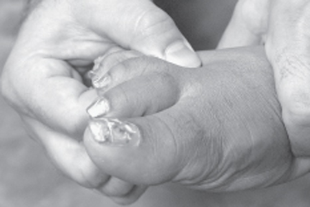
48 Morton Neuroma A 45-year-old, overweight woman presented to her primary care doctor with a complaint of increasing pain in her distal right foot. She described the pain as if there was “hot gravel digging into the sole” of her foot. This pain was made worse by weight bearing. She obtained significant relief by removing her shoes, rubbing the sole of her foot, and flexing her toes. On physical exam the patient had exquisite point tenderness on the plantar surface of her foot ˜2 cm proximal to the apex of her third web space. Her motor and sensory exams were normal. The diagnosis of Morton neuroma was made. Her pain failed to resolve after 4 weeks of Neurontin (Pfizer, Inc., New York, NY) therapy and use of an orthotic shoe. She subsequently underwent a resection of the Morton neuroma through a dorsal approach under local anesthesia and posterior tibial nerve block. Upon resuming full weight bearing 1 week later she had no point tenderness in her foot and noted a small area of decreased sensation along the inside of her third web space. Morton neuroma (plantar interdigital neuroma) Morton neuroma is a benign, painful enlargement of one of the common digital nerves of the foot. However, the lesion almost always afflicts the third common digital nerve. Histopathologically, the lesion is not a true neuroma. Rather, the most frequently reported pathological findings are fibrosis and axonal degeneration. These findings suggest that the etiology of this condition is more akin to a compressive entrapment phenomenon. The third common digital nerve is exceedingly more vulnerable to this disorder than the other common digital nerves. This particular vulnerability is likely related to the unique anatomy of the third common digital nerve. The third is the only common digital nerve to receive a contribution from both the lateral and the medial plantar nerves. As these nerves course anteriorly into the forefoot, the medial plantar nerve is immediately medial to and the lateral plantar nerve is deep to the flexor digitorum brevis muscle belly. This muscle belly forms the “brevis sling,” which is tightened with foot dorsiflexion and relaxed with toe flexion. When the brevis sling is tightened during dorsiflexion or toe extension the lateral and medial plantar nerve contributions to the third common digital nerve are stretched. This then places a stretch on the common digital nerve, which pushes the nerve against the plantar surface of the taught and firm deep transverse intermetatarsal ligament that lies immediately dorsal to the nerve. Although this mechanism has not been proven in a rigorous fashion, it is plausible and well accounts for the repetitive traumatic changes that are seen in the pathological studies of excised interdigital “neuromas.” A Morton neuroma produces pain in the forefoot. The pain is almost always localized to the plantar surface of the forefoot approximately 2 cm proximal to the third web space. The second web space is the next most common site of pain. The pain may radiate distally into the inner surface of the distal toes or proximally in the heel of the foot. The pain is exacerbated by weight bearing and by tight-fitting shoes. Classically, removing the shoes and massaging the foot relieve the pain. Toe plantarflexion also relieves the pain. The syndrome is much more common in women than in men. Patients will typically present between the fourth and sixth decades. A history of wearing high-heeled shoes is frequently present. The pain generally develops insidiously and symptoms are often present for several months before medical attention is sought. An acute onset or an exacerbation following seemingly mild foot trauma may prompt earlier medical attention. Although the presentation described here is virtually pathognomonic for Morton neuroma, there are other causes of pain in the plantar forefoot. The most common would be metatarsal bursitis or a metatarsal stress fracture. Other potential causes would include diabetic neuropathy, tarsal tunnel syndrome, rheumatoid arthritis, or localized vasculitis. Weight-bearing radiographs should be obtained to rule out metatarsal fractures as a potential cause. Nerve conduction studies should be considered in patients whose medical history or presentation suggests the possibility of a metabolic neuropathy or tarsal tunnel syndrome. The diagnosis of Morton neuroma can usually be made by the characteristic presenting complaints obtained from the patient’s history. The examiner should localize the area of tenderness, which should be located ˜2 cm proximal to the third or second web space. The patient’s pain should be reproduced by the examiner squeezing the five metatarsal heads together with one hand while simultaneously compressing the third web space with the thumb and forefinger of the other hand from both the plantar and dorsal directions (Fig. 48–1). Placing the foot in dorsiflexion and the toes in extension will also likely exacerbate the patient’s pain. The Morton neuroma may be large enough to present as a palpable mass at the site of point tenderness, but this is not required for the diagnosis. Sensation is usually preserved but some light touch hypoesthesia may be present on the toe surfaces of the involved web space. A Tinel sign may be present at the site of point tenderness. Motor weakness should not be encountered. A weight-bearing foot radiograph should be performed to evaluate for any bony abnormality that may cause foot pain or complicate surgical treatment of the Morton neuroma. Magnetic resonance imaging (MRI) of the affected foot may detect a Morton neuroma. High-resolution ultrasonography appears to be the imaging study of choice if the sonographers have experience evaluating this disorder. Sonography data suggest that a neuroma becomes symptomatic when it approaches 5 mm in diameter. The normal common interdigital nerve diameter is ˜2 mm. Electrodiagnostic testing is generally not helpful in the evaluation of Morton neuroma. It is difficult to localize a specific common interdigital nerve with a needle electrode to measure sensory conduction. Nerve conduction studies may be helpful to rule out other potential causes of foot pain such as diabetic neuropathy and tarsal tunnel syndrome. The initial intervention with Morton neuroma should be an attempt to alleviate aggravating factors. This attempt should include the avoidance of high-heeled shoes and other narrow shoes that compress the metatarsal heads. An orthotic shoe with padding that cushions the second, third, and fourth metatarsals may also provide some pain relief. The avoidance of weight bearing is not a practical or long-term remedy. An injection of steroids along with a local anesthetic may provide short-term relief and less frequently may result in long-term resolution of the pain. The injection solution should be delivered from both the dorsal and the plantar surfaces of the forefoot and directed toward the point of maximal tenderness. Unfortunately, these conservative measures often fail to provide satisfactory pain resolution, and surgical intervention must be considered in many cases.
 Case Presentation
 Diagnosis
 Anatomy
 Characteristic Clinical Presentation
 Differential Diagnosis
 Diagnostic Tests
 Management Options

Stay updated, free articles. Join our Telegram channel

Full access? Get Clinical Tree


