CHAPTER 392 Natural History of Cavernous Malformations
Before the availability of modern imaging technology, cavernous malformations were considered rare lesions. In 1976, Voigt and Yasargil described their clinical experience with one patient and thoroughly reviewed the world literature, finding only 126 reported cases.1 Soon after the publication of this article, computed tomography (CT) became widely available; however, CT lacked both the sensitivity and specificity for identifying cavernous malformations. Only partially calcified or recently hemorrhagic lesions could be readily visualized, and diagnosis still required pathologic confirmation.
The subsequent introduction of magnetic resonance imaging (MRI) in the mid-1980s revolutionized our understanding of these lesions. The MRI characteristics of cavernous malformations are sufficiently unique to allow diagnosis of the majority of these lesions on the basis of the MRI findings alone (Fig. 392-1).2–6 The widespread availability of MRI has increased the recognition of cavernous malformations in both symptomatic and asymptomatic patients and the number of requests for neurosurgical consultation. Appropriate management of these patients requires a thorough understanding of the epidemiology and natural history of these lesions. The goals of this chapter are to provide the reader with an in-depth review of the available literature on this topic and to examine the implications related to the treatment of these patients.
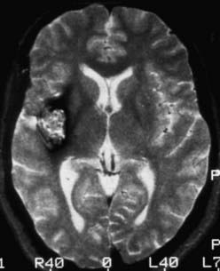
FIGURE 392-1 Axial T2-weighted magnetic resonance image demonstrating the classic appearance of a cavernous malformation. The core of the lesion has a reticulated “salt and pepper” pattern and is surrounded by a halo of low signal intensity. Histologically, these lesions are composed of loculated areas of hemorrhage and thrombosis of various age surrounded by gliotic, hemosiderin-stained brain tissue. Note the absence of mass effect and edema. This pattern of hemorrhage (type II) is pathognomonic for a cavernous malformation. See Table 392-3 for the classification of lesion types.
Epidemiology
Cavernous malformations are more common than generally suspected. Postmortem studies from the 1980s (Table 392-1) demonstrated that cavernous malformations affect 0.37% to 0.5% of the population.7,8 Remarkably similar results were reported by two groups reviewing more than 22,000 MRI examinations, for which incidence rates of 0.4% to 0.5% were calculated.9,10 Based on these studies, it is estimated that 1 in every 200 to 250 people, or approximately 30 million individuals worldwide, are affected by cavernous malformations.
TABLE 392-1 Incidence of Cavernous Malformations
| REFERENCE | NO. PATIENTS STUDIED | NO. LESIONS/INCIDENCE RATE OF CAVERNOUS MALFORMATIONS |
|---|---|---|
| McCormick,7 1984 | 5,734 | 19/0.34% |
| Otten et al.,8 1989 | 24,535 | 131/0.53% |
| del Curling et al.,9 1991 | 8,131 | 32/0.39% |
| Robinson et al.,10 1991 | 14,035 | 66/0.47% |
| Total | 52,435 | 248/0.47% |
Cavernous malformations occur in two forms: spontaneous and familial. The spontaneous form occurs as an isolated event, most commonly with a single lesion. The familial form is characterized by multiple lesions (Fig. 392-2) and an autosomal dominant mode of inheritance.11–14 At least three distinct genetic loci have been identified with this form of the disease.15–17 A detailed review of the genetics of familial cavernous malformations is beyond the scope of this chapter; interested readers are referred to Chapter 393.
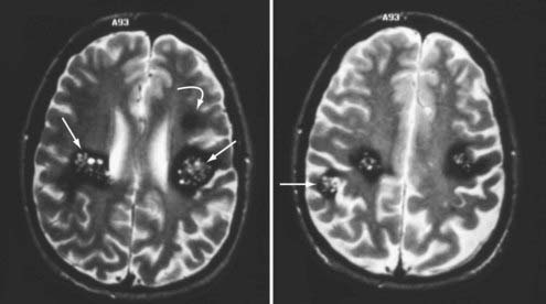
FIGURE 392-2 Axial T2-weighted magnetic resonance images in a patient with a new onset of seizures and familial cavernous malformations. Four distinct lesions are seen at these two levels, including three type II lesions (straight arrows) and one type III lesion (curved arrow). A fifth lesion was identified in the cerebellum. Multiple lesions and a family history of seizures are characteristic of the familial form of this disease. See Table 392-3 for the classification of lesion types.
The presence of three or more lesions and a family history of seizures are essentially pathognomonic for the familial form of this disease. In a recent series of 132 patients with familial cavernous malformations, Denier and coworkers reported that 80% had multiple lesions detected on T2-weighted MRI and 90% had multiple lesions on gradient-echo images, with an average of 5 lesions per patient on T2-weighted images and 20 per patient on gradient-echo sequences.18 A thorough family history is indicated for any patient with multiple cavernous malformations.
Cavernous malformations occur throughout the central nervous system in rough proportion to the volume of the various compartments: supratentorial, 80%; brainstem and basal ganglia, 15%; and spinal cord, 5%. They have been well described in infants and children but are seldom symptomatic until the second and third decades of life. Large population studies have demonstrated that cavernous malformations occur with equal frequency in both sexes.9,10 Some data suggest that women may more commonly be seen initially with symptomatic hemorrhage, but the evidence is far from conclusive. The issues of gender and pregnancy are covered later in this chapter.
Not all patients with cavernous malformations become clinically symptomatic. Twenty percent to 30% of sporadic lesions are incidental findings discovered during evaluation for headache or other unrelated symptoms.9,10 Approximately 40% of patients with the familial form of the disease remain asymptomatic despite the presence of multiple lesions.13,19
Clinical Findings
Evidence of previous hemorrhage is a constant feature of cavernous malformations regardless of whether the lesions are symptomatic. Hemorrhages of various ages, combined with the deposition of hemosiderin in cerebral tissue surrounding the cavernous malformation, produce the unique MRI characteristics of these lesions (see Figs. 392-1 and 392-2). Increases in size may result from repeated small hemorrhages within the lesion and from spontaneous thrombosis of the blood-filled caverns. Organization and endothelialization within these hemorrhagic/thrombotic cavities create the potential for further growth. Rarely, these lesions rupture outside their capsule and produce “overt” hemorrhage into the surrounding brain tissue (Figs. 392-3 and 392-4). Because cavernous malformations are low-flow, low-pressure lesions, hemorrhage (even “overt” hemorrhage outside the lesion) usually displaces and compresses adjacent neural tissue rather than destroying it.
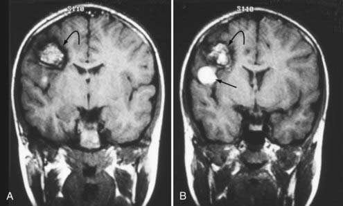
FIGURE 392-3 A and B, Coronal T1-weighted magnetic resonance images in a 10-year-old boy with a cavernous malformation (curved arrows). His past history was remarkable for an episode of seizure activity at 4 years of age and a negative computed tomographic study. The patient had a sudden onset of mild weakness and decreased sensation in his left upper extremity. Note the focal area of subacute blood (straight arrow) extending outside the capsule of the lesion and producing “overt” extralesional (type IA) hemorrhage. See Table 392-3 for the classification of lesion types.
(From Zabramski JM. Cavernous malformations. In: Aminoff M, Daroff RB, eds. Encyclopedia of the Neurological Sciences. San Diego, CA: Academic Press; 2003. Used with permission.)
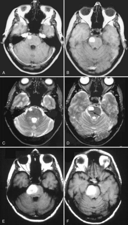
FIGURE 392-4 This 38-year-old woman was seen at an outside emergency department with complaints of severe headache and nausea. A basic head computed tomographic scan revealed a subtle area of increased density in the right pons. T1-weighted (A and B) and T2-weighted (C and D) magnetic resonance imaging (MRI) revealed a 1.6-cm area of subacute blood consistent with recent “overt” extralesional (type IA) hemorrhage from a small cavernous malformation (arrow), which is best seen on the T2-weighted image (D). The patient was seen in neurosurgical consultation approximately 1 month after this episode when all symptoms had resolved. She initially declined surgical intervention. Two months later, she had an acute onset of left-sided weakness and hemisensory deficits. Repeated T1-weighted MRI (E and F) revealed a dramatic increase in the size of the lesion, which was composed almost entirely of acute and subacute blood. Surgical pathologic evaluation confirmed the diagnosis of cavernous malformation. After resection of this lesion, she made a complete recovery. See Table 392-3 for the classification of lesion types.
(C and D, From Zabramski JM, Spetzler RF. Management of brainstem cavernous malformations. In: Batjer HH, Loftus CM, eds. Textbook of Neurological Surgery. Philadelphia: Lippincott, Williams & Wilkins; 2003. Used with permission; E and F, from Robinson JR, Zabramski JM. Cavernous malformations of the cervicomedullary junction. In: Dickman CA, Spetzler RF, Sonntag VKH, eds. Surgery of the Craniovertebral Junction. New York: Thieme; 1998.)
Supratentorial Lesions
Seizures are the most common manifestation of supratentorial cavernous malformations and account for 40% to 80% of the initial symptoms.9,10,12,13,20–22 Despite the prevalence of seizures in this population, remarkably few data are available regarding the annual risk for the onset of seizures. Only four studies directly addressed this issue and reported rates for new onset of seizures of 1.4% to 2.4% per person-year.9,19,23,24 The onset or exacerbation of seizure activity in these patients is often associated with MRI evidence of acute/subacute hemorrhage (Fig. 392-5). The exact mechanism that leads to the seizure activity in these lesions is unknown. Cavernous malformations do not typically contain neuronal tissue and are therefore not intrinsically epileptogenic. They induce seizures through their effect on surrounding brain tissue. Such effects may include focal gliosis, hemosiderin deposition, and cellular and humoral inflammatory responses. Pathologically, cavernous malformations are surrounded by a yellow-brown–stained border that is heavily infiltrated with hemosiderin-laden macrophages and iron (Fig. 392-6). Iron is a well-known epileptogenic material used to induce seizures in laboratory models of epilepsy.25,26 Focal neurologic deficits secondary to mass effect are rarely associated with supratentorial lesions unless the lesion is located in the basal ganglia or thalamus.
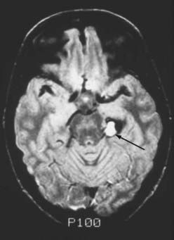
FIGURE 392-5 Intermediate-weighted, axial, spin echo magnetic resonance image in a 23-year-old man with acute exacerbation of temporal lobe seizure activity. Note the focal area of high-signal intensity (arrow) compatible with subacute hemorrhage in the region of the left hippocampus. A ring of low signal intensity surrounds the entire lesion, consistent with an intralesional (type IB) hemorrhage. Surgical pathologic evaluation confirmed the diagnosis of cavernous malformation. See Table 392-3 for classification of lesion types.
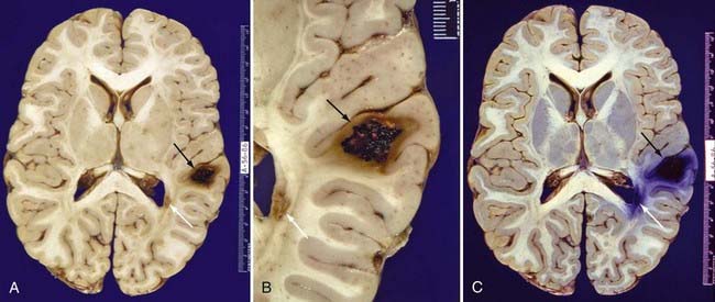
FIGURE 392-6 Whole-brain sections from a 26-year-old Hispanic man who died of septic complications. A, An unstained formalin-fixed section reveals a type II cavernous malformation (black arrow; see Table 392-3 for definition) in the right posterior frontal lobe and a small type III cavernous malformation (white arrow; see Table 392-3 for definition) adjacent to the right occipital horn. B, Close-up view of the same lesions. C, The same section stained with Prussian blue for iron emphasizes the extent of hemosiderin and iron deposition. The hemosiderin and iron surrounding these lesions are responsible for the areas of low signal intensity (known as blooming artifact) seen on T2-weighted and gradient-echo magnetic resonance images.
(A and B, From Zabramski JM. Cavernous malformations. In: Aminoff M, Daroff RB, eds. Encyclopedia of the Neurological Sciences. San Diego, CA: Academic Press; 2003. Used with permission; C, used with permission from Barrow Neurological Institute.)
Brainstem Lesions
The sudden onset of focal neurological deficits is the most frequent finding in patients with brainstem cavernous malformations.27–30 Porter and coauthors reported 100 surgical cases of brainstem cavernous malformations. In their series, 97% of patients had focal neurological deficits from hemorrhage.30 The development of symptoms in patients with hemorrhage from brainstem cavernous malformations is characteristically acute and maximal at onset (see Fig. 392-4). However, the neurological deficits from the first episode of clinically symptomatic hemorrhage tend to resolve completely as the hemorrhage is organized and absorbed. In contrast, recurrent episodes of hemorrhage are apt to be associated with progressively more severe deficits and an increased risk for permanent neurological impairment (see Fig. 392-4). This stuttering clinical course with improvement of symptoms between episodes may lead to a misdiagnosis of multiple sclerosis if the evaluation does not include MRI. Occasionally, large brainstem lesions develop that are associated with only minimal deficits, particularly in the pons, where the mass can gradually displace the densely packed ascending and descending fiber tracts (Fig. 392-7). Death is rare without multiple episodes of symptomatic hemorrhage.
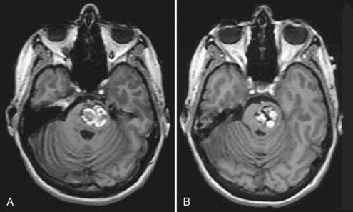
FIGURE 392-7 A and B, T1-weighted magnetic resonance images in a 39-year-old woman with a history of an acute onset of mild right-sided weakness that had completely resolved over a period of 3 to 4 weeks. The hyperintense regions represent areas of subacute hemorrhage within the more chronic matrix of this pontine cavernous malformation. This lesion reached considerable size with only minimal deficits by gradually displacing the dense fiber tracts within the pons. Compare this pattern with the “overt” pontine hemorrhage sustained by the patient shown in Figure 392-4.
Spinal Cord Lesions
Ogilvy and associates described four types of manifestations in patients with symptomatic intramedullary spinal cord lesions31: (1) acute onset of symptoms with a rapid decline, (2) acute onset of mild symptoms with a subsequent gradual decline, (3) acute episodes of stepwise neurological deterioration, and (4) slow progression of neurological deterioration. Sandalcioglu and colleagues noted that the first three of these categories all seem to be related to an acute event leading to neurological deficits of varying severity and course.32 Consequently, they recommended that patients be separated into only two subgroups. One group is typified by major hemorrhage and the sudden onset of symptoms and neurologic deficits, the gravity of which is related to the exact location of the lesion (level and position within the cord) and the extent of hemorrhage (intralesional or extralesional volume, or both). The second group is characterized by slowly progressive myelopathic or radicular symptoms (or both), probably related to minor bleeding episodes and gradual growth of the cavernous malformation. Combinations of these two manifestations occur frequently.
Both radicular and myelopathic symptoms are common at initial evaluation. Pain is a significant component in 40% to 50% of patients with spinal cord lesions31,33–37 and was the only symptom in approximately 25% of patients in a series from our institution.38
It is important to note that patients with spinal cord cavernous malformations appear to be at significantly increased risk of harboring intracranial lesions. Vishteh and colleagues reported that 47% of patients with symptomatic spinal cord lesions who undergo MRI of the complete neuraxis will be found to have intracranial lesions.39 Cohen-Gadol and coworkers recently confirmed this finding and reported a 40% incidence of intracranial lesions in a series of 33 patients with spinal cord cavernous malformations who underwent both cranial and spinal imaging studies.40 These reports suggest that it may be prudent to perform MRI of the brain in this population.
Natural History
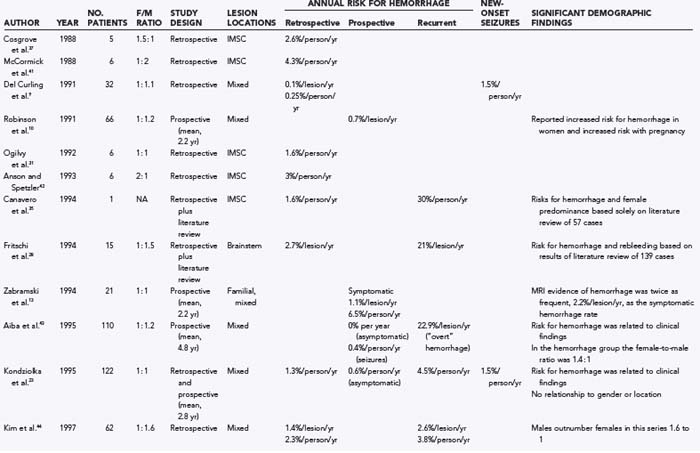
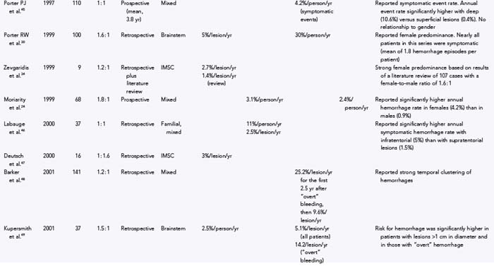
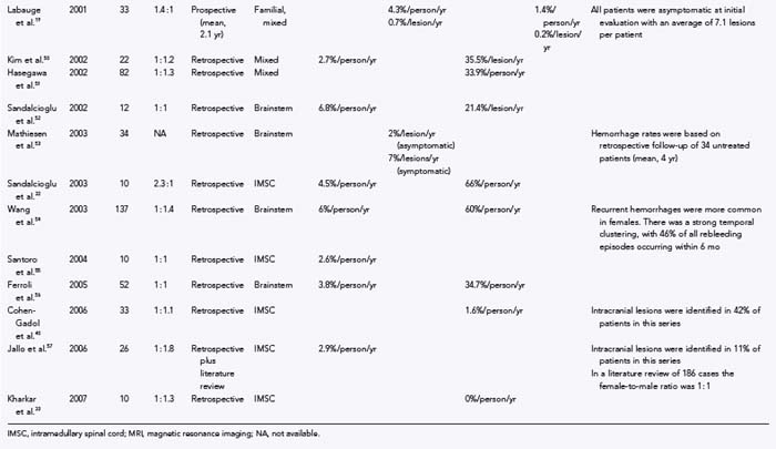
Numerous studies have been published on the natural history of cavernous malformations (Table 392-2).* Hemorrhage rates vary widely from series to series depending on the authors’ definition of hemorrhage and the population being studied. Not surprisingly, hemorrhage rates tend to be higher in surgical series because patients with symptomatic lesions are more likely to be referred for neurosurgical intervention. In addition, there are essentially four different methods of calculating hemorrhage rates, including retrospective and prospective methods, either of which can be reported as risk for hemorrhage per patient or per lesion. The retrospective method assumes that all lesions have been present since birth. Using this assumption, Del Curling and collaborators calculated an annual hemorrhage rate of 0.25% per patient.9 Kondziolka and coauthors reported a rate of 1.3% per patient per year,23 and Kim and associates calculated a rate of 2.3% per patient per year.44 This method of calculation, which depends on the patient’s recall to define episodes of hemorrhage and assumes that all lesions are present from birth, is likely to underestimate the actual risk of significant bleeding. Once considered congenital in origin, there is increasing evidence that new lesions may appear de novo in both the sporadic and familial forms of the disease.13,14,58–68
Stay updated, free articles. Join our Telegram channel

Full access? Get Clinical Tree


