Neurocutaneous Syndromes


Neurocutaneous Syndromes


Marc C. Patterson


INTRODUCTION

Several genetic diseases involve both the skin and nervous system. These are called neurocutaneous disorders or neuroectodermatoses. In the past, they were referred to as the phakomatoses (phakos is the Greek word for lentil, flat plate, or spot).

Any part of the central nervous system (CNS) or peripheral nervous system can be affected by these hereditary diseases; their expressivity is quite variable both between and within kindreds.


NEUROFIBROMATOSIS


INTRODUCTION

Although neurofibromatosis (NF) was first described at least as early as the 17th century and a comprehensive clinical review was published in 1849, the clinical and pathologic phenotypes were first linked by von Recklinghausen in 1882. NF is one of the most common single-gene disorders of the CNS. Cardinal features include multiple cutaneous hyperpigmented marks (café au lait spots); multiple neurofibromas; and lesions in bone, CNS, peripheral nervous system, or other organs. The best characterized forms are neurofibromatosis type 1 (NF-1; von Recklinghausen disease or peripheral NF [MIM 162200]), neurofibromatosis type 2 (NF-2; central NF or the bilateral acoustic neuroma syndrome [MIM 101000]) and schwannomatosis, a phenotype characterized by the presence of multiple schwannomas, associated with intractable pain, without vestibular involvement. The three conditions differ in genetics, pathogenesis, and clinically.


EPIDEMIOLOGY

Both sexes are affected equally, and the condition is found worldwide in all racial and ethnic groups. Incidence figures must be a minimal estimate, because mild cases are often unrecognized.


MOLECULAR GENETICS AND PATHOBIOLOGY

NF-1, NF-2, and schwannomatosis are autosomal dominant conditions; penetrance of NF-1 is almost 100%, but expressivity varies. Mutations account for 50% of new cases. The NF-1 gene has been mapped to chromosome 17q11.2. The gene product is called neurofibromin, a tumor suppressor gene that acts through the RAS/MAPK pathway. Translocations, large megabase deletions, large internal deletions, small rearrangements, and point mutations all occur in the NF-1 gene. Although the genotypes differ, the phenotypes are indistinguishable. The marked clinical variability within families having an identical NF-1 gene mutation equals the variability among families with different NF-1 gene mutations. The exception occurs with large megabase deletions associated with cognitive dysfunction and more severe clinical manifestations generally. Mutation inactivates the gene, and, by analogy with other oncogenes, loss of the allelic gene later in life could result in tumor formation. It is not known how the other manifestations of the disease arise. Several potential genetic modifiers in associated signalling pathways have been identified in screening studies.

The NF-2 gene maps to chromosome 22q12. The gene product is similar to that of the moezin-ezrin-radixin-like protein gene; for this reason, it is called merlin. Deletions of the gene have been found in schwannoma and meningioma cells, the major tumors in NF-2 patients. NF-2 inhibits the Rac/CDC42-dependent Ser/Thr kinase PAK1, which activates both Ras transformation and NF-1 through two separate domains. Specific inhibitors of PAK1 selectively inhibit the growth of NF-2-deficient cancer cells, suggesting that PAK1 is essential for the malignant growth of NF-2-deficient cells. Inhibitors of PAK1 might have a future role as therapies for these tumors.

Schwannomatosis has been linked to mutations in three genes: SMARCB1, LZTR1, and COQ6. The SMARCB1 gene product is a subunit of the SWI/hSHNF chromatin remodeling complex. Mutations in this gene account for about half of familial schwannomatosis but only about 10% of sporadic cases. LZTR1 encodes a protein that acts as a tumor suppressor; COQ6 mutations impair the synthesis of coenzyme Q10.


NEUROPATHOLOGY

Dysplasia, hyperplasia, and neoplasia of neural-supporting tissues involves the central, peripheral, and autonomic nervous systems. Visceral manifestations result from hyperplasia of the autonomic ganglia and nerves within the organ. Dysplastic and neoplastic changes also affect skin, bone, endocrine glands, and blood vessels. Developmental anomalies include thoracic meningocele and syringomyelia. Patients affected by NF-1 are more likely than others to have neoplastic disorders, including neuroblastoma, Wilms tumor, leukemia, pheochromocytoma, and sarcomas.

Neoplasms involving the peripheral nervous system and spinal nerve roots include schwannomas and neurofibromas. Intramedullary spinal cord tumors include ependymomas (especially of the conus medullaris and filum terminale) and astrocytomas (although some of these may actually be ependymomas when electron microscopy is used); gangliomas are rare. The most common intracranial tumors are hemispheric astrocytomas of any histologic grade from benign to highly malignant; posterior fossa tumors are rare. Pilocytic astrocytic gliomas of the optic nerve and optic chiasm are also characteristic and generally progress very slowly. Bilateral acoustic neuromas and solitary or multicentric meningiomas occur in adults with NF-2.


CLINICAL MANIFESTATIONS

There are at least four forms of NF. Peripheral NF (NF-1) as described by von Recklinghausen is most common. Central NF (NF-2) is manifested by bilateral acoustic neuromas with mild cutaneous changes. Segmental NF arises from mosaic-type microdeletions in NF-1; it is characterized by café au lait spots and neurofibromas that are limited, usually affecting an upper body segment. The lesions extend to the midline and include the ipsilateral arm but
spare the head and neck. Cutaneous NF is limited to pigmentary changes; there are numerous café au lait spots but no other clinical manifestations. One percent to 4% of patients with a mild NF phenotype (café au lait spots only) have a related disorder, Legius syndrome, which is associated with mutations in the SPRED1 gene.

NF-1-Noonan syndrome has the clinical manifestations of NF-1 coupled with a Noonan syndrome phenotype. The clinical features of the Noonan syndrome include ocular hypertelorism, downward-slanting palpebral fissures, webbed neck, low-set ears, and pulmonic stenosis.

NF-1 has protean and progressive manifestations. Families and physicians often anticipate a fate like that of Joseph Merrick (the Elephant Man, who almost certainly had the Proteus syndrome, not NF-1). In reality, many patients with the disease are functionally indistinguishable from normal. Often, they have only cutaneous lesions and are not diagnosed until they see a physician because of a learning disability, scoliosis, or another problem.


Cutaneous Manifestations

The café au lait macule is the pathognomonic lesion, being present in almost all patients (Figs. 140.1 and 140.2). Six or more café au lait spots larger than 5 mm in diameter before puberty and greater than 15 mm in diameter after puberty are diagnostic. The spots are usually present at birth but may not appear until age 1 or 2 years. Increasing in both size and number during the first decade of life, the macules tend to be less evident after the second decade because they blend into the surrounding hyperpigmented skin. These discrete, tan macules involve the trunk and limbs in a random fashion but tend to spare the face.

Other cutaneous manifestations may include freckles over the entire body, but freckles usually involve the axilla and other intertriginous areas. Larger, darker hyperpigmented lesions are often associated with an underlying plexiform neurofibroma if this involves the midline, it may indicate the presence of a spinal cord tumor.


Ocular Findings

Pigmented iris hamartomas (Lisch nodules) (Fig. 140.3) are pathognomonic and consist of small translucent yellow or brown elevations on slit lamp examination. The nodules increase in number with age and are present in almost all patients older than 20 years.


FIGURE 140.1 Café au lait patches and neurofibromas on the back. (Courtesy of Suresh Kotagal, MD.)


FIGURE 140.2 Cafe-au-lait patches on the abdomen. (Courtesy of Marc C. Patterson, MD.)


Skeletal and Limb Manifestations

Skeletal anomalies characteristic of NF include (1) unilateral defects in the posterosuperior wall of the orbit, with pulsating exophthalmos, usually associated with dural ectasia; (2) a defect in the lambdoid with underdevelopment of the ipsilateral mastoid; (3) dural ectasia with enlargement of the spinal canal and scalloping of the posterior portions of the vertebral bodies (also seen in connective tissue disorders such as Marfan and Ehlers-Danlos syndromes); (4) scoliosis, affecting between 21% and 49% of patients with NF-1; (5) pseudarthrosis, especially involving the tibia (5% of cases) and radius; (6) “twisted ribbon” rib deformities; and (7) enlargement of long bones.


Miscellaneous Features

Pheochromocytomas occur in 0.1% to 5.7% of patients with NF-1; they are rare in children. Hypertension may be due to a pheochromocytoma (20% to 50% of cases) or a neurofibroma of a renal artery. Malignant tumors occurring less frequently in NF include sarcoma, leukemia, Wilms tumor, ganglioglioma, and neuroblastoma. Medullary thyroid carcinoma and hyperparathyroidism are rare. Precocious puberty and, less commonly, sexual infantilism result from involvement of the hypothalamus by glioma or hamartoma. Cystic lesions, malignancy, and interstitial pneumonia are pulmonary complications.


TUBEROUS SCLEROSIS COMPLEX


INTRODUCTION

Tuberous sclerosis complex (TSC) was first described by von Recklinghausen in 1863. In 1880, Bourneville coined the term sclérose tubéreuse for the potato-like lesions in the brain. In 1890, Pringle described the facial nevi or adenoma sebaceum. Vogt later emphasized the classic triad of seizures, intellectual disability, and adenoma sebaceum. TSC (MIM 191400) is a progressive genetic disorder characterized by the development in early life of hamartomas, malformations, and congenital tumors of the CNS, skin, and viscera.


EPIDEMIOLOGY

Incidence figures are considered minimal because milder varieties are often unrecognized. Clinical surveys suggest a prevalence between 1 in 10,000 and 1 in 170,000. Pulmonary lymphangioleiomyomatosis, progressive and often fatal, occurs almost exclusively in young women.


GENETICS

Tuberous sclerosis is an autosomal dominant trait, with a high incidence of sporadic cases and protean clinical expression. This variability is attributed to modifier genes. The defective genes map to chromosome 9q34 (TSC1) and chromosome 16p13.3 (TSC2). TSC1 or TSC2 is mutated in 75% to 85% of TSC patients; no mutation is found in 15%. Hamartin is the gene product for TSC1, and tuberin is the gene product for TSC2. Hamartin and tuberin form heterodimers that inhibit the mammalian target of rapamycin (mTOR); phosphorylation is an important aspect of this complex process, which involves over 50 interacting proteins.


PATHOLOGY AND PATHOBIOLOGY

The pathologic changes are widespread and include lesions in the nervous system, skin, bones, retina, kidney, lungs, and other viscera. Multiple small nodules often line the ventricles.

TSC is characterized by the presence of hamartias and hamartomas. Hamartias (from the Greek for “flaw”) are malformations in which cells native to a tissue display abnormal architecture and morphology. These lesions do not grow disproportionately for the tissue or organ in which they are found. Hamartomas have the same characteristics but grow excessively for their site of origin. This old concept is valuable in recognizing the proliferative potential of lesions found in TSC. Thus, cortical tubers are hamartias and do not grow excessively, whereas angiomyolipomas are hamartomas that grow disproportionately and may produce symptoms as a consequence.

The brain is usually normal in size, but variable numbers of hard nodules occur on the surface of the cortex. These nodules are smooth, round, or polygonal and project slightly above the surface of the neighboring cortex. They are white, firm to the touch, and of various sizes. Some involve only a small portion of one convolution; others encompass the convolutions of one whole lobe or a major portion of a hemisphere. In addition, there may be developmental anomalies of the cortical convolutions in the form of pachygyria or microgyria. On sectioning of the hemispheres, sclerotic nodules may be found in the subcortical gray matter, the white matter, and the basal ganglia. The lining of the lateral ventricles is frequently the site of numerous small nodules that project into the ventricular cavity. Sclerotic nodules are less frequently found in the cerebellum. The brain stem and spinal cord are rarely involved.

Histologically, the nodules are characterized by a cluster of atypical glial cells in the center and giant cells in the periphery. Calcifications are relatively frequent. Other features include heterotopia, vascular hyperplasia (sometimes with actual angiomatous malformations), disturbances in the cortical architecture, and, occasionally, development of subependymal giant cell astrocytomas (SEGAs). Intracranial giant aneurysm and arterial ectasia are uncommon findings.

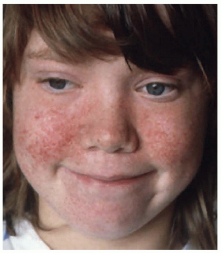
The skin lesions are multiform and include the characteristic facial angiofibromas and fibrous plaques, typically localized to the frontal area. The facial lesions are not adenomas of the sebaceous glands but small hamartomas arising from nerve elements of the skin combined with hyperplasia of connective tissue and blood vessels (angiofibromas) (Fig. 140.4). In late childhood, lesions similar to those on the face are found around or underneath the nails (periungual or 
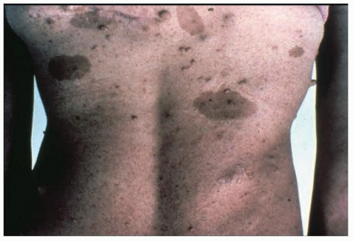
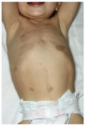
subungual fibromas). Circumscribed areas of
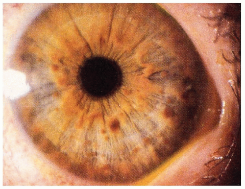
hypomelanosis or depigmented nevi are common in tuberous sclerosis and are often found in infants. Although these depigmented nevi are less specific than the angiofibromas, they are important signals of the diagnosis in infants with seizures. Histologically, the skin appears normal except for the loss of melanin, but ultrastructural studies show that melanosomes are small and have reduced content of melanin. The retinal lesions are small congenital tumors composed of glia, ganglion cells, or fibroblasts. Glioma of the optic nerve has been reported.


FIGURE 140.4 Facial angiofibromas.

Other lesions include cardiac rhabdomyoma; renal angiomyolipoma, renal cysts, and, rarely, renal carcinoma; cystic disease of the lungs and pulmonary lymphangioleiomyomatosis; hepatic angiomas and hamartomas; skeletal abnormalities with localized areas of osteosclerosis in the calvarium, spine, pelvis, and limbs; cystic defects involving the phalanges; and periosteal new bone formation confined to the metacarpals and metatarsals.


CLINICAL MANIFESTATIONS

The cardinal features of tuberous sclerosis are skin lesions, convulsive seizures, and intellectual disability. The disease is characterized by variable expressivity of the clinical manifestations and is often age related: the symptomatic neonate with cardiac rhabdomyoma and heart failure; the infant with hypomelanotic macules and infantile spasms; preschool and school-age children with angiofibromas, developmental delay, learning disability or intellectual disability, and seizures; and adults with dermatologic lesions, subungual fibromas, seizures, and often intellectual disability.

Only gold members can continue reading. Log In or Register to continue

Stay updated, free articles. Join our Telegram channel

Jul 27, 2016 | Posted by in NEUROLOGY | Comments Off on Neurocutaneous Syndromes

Full access? Get Clinical Tree

Get Clinical Tree app for offline access