Pain, Numbness, and Paresthesias


Pain, Numbness, and Paresthesias


Comana M. Cioroiu


INTRODUCTION

For the general neurologist, the evaluation of a patient with complaints of numbness can be a daunting one. Being the most subjective of neurologic complaints, numbness is often a difficult sensation for the patient to explain and often all the more difficult for the clinician to appreciate on examination.


MANAGEMENT STRATEGY

As with all complaints in medicine, the first step is to obtain a detailed history. The first question should be one aimed at best characterizing the sensation (or lack thereof) described by the patient. It is important to differentiate between numbness as a loss of sensation as opposed to the presence of an abnormal sensation. At times, patients may also use the term numbness to describe muscle weakness, and this is important to keep in mind during one’s examination.


NEUROPATHIC PAIN

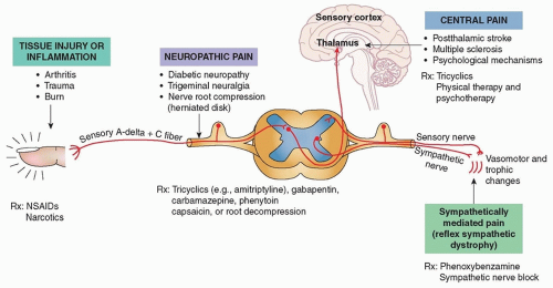
Neuropathic pain (see Chapter 57) is a category of pain specific to that caused by nerve injury and is often described as painful paresthesias, associated with a sensation of burning or radiating pain as can be seen in cases of peripheral neuropathy or radiculopathy (Fig. 8.1). In such cases, patients may exhibit altered sensation or abnormally increased sensations— hyperesthesia refers to increased sensation, whereas dysesthesia refers to an evoked unpleasant or painful sensation.

An exaggerated response to stimuli that normally evoke pain is known as hyperalgesia, whereas an exaggerated response to stimuli that should not normally invoke pain is referred to as allodynia. These abnormal sensations can be seen with various nerve injuries, both at the central and peripheral level. Conversely, hypoesthesia refers to the diminished perception of pain, anesthesia to the inability to perceive pain, and analgesia to the inability to feel pain.

Neuropathic pain must be differentiated from somatic pain caused by pathology in bones, ligaments, muscles, and other soft tissues.

Complex regional pain syndrome (CRPS; also known as reflex sympathetic dystrophy) is a chronic disease characterized by severe neuropathic pain, skin changes, and swelling in one limb. It may occur in the absence of a known nerve injury (type 1) or in the
setting of known nerve injury (type 2). Diagnosis and treatment of CRPS is further discussed in Chapter 56.


TABLE 8.1 Patterns of Sensory Loss


Localization


Sensory Pattern


Associated Findings


Cortical


  • Contralateral sensory loss


  • Cortical sensory loss (i.e., neglect, astereognosis, agraphesthesia)


  • Visuospatial deficits


Brain stem


  • Contralateral sensory loss of arm and leg with ipsilateral loss in face


  • Cranial neuropathies (i.e., ophthalmoparesis, facial weakness)


  • Ataxia


  • Contralateral weakness


  • Hyperreflexia


Spinal cord


  • Complete transverse lesion: loss of all sensory modalities below the level of the lesion


  • Central cord lesion (i.e., syrinx): loss of pain and temperature below the level of the lesion


  • Anterior cord lesion: contralateral loss of pain and temperature sensation below level of the lesion


  • Cord hemisection (Brown-Séquard syndrome): ipsilateral loss of vibration and joint position below the level of the lesion and contralateral loss of pain and temperature two or three segments below the level of the lesion


  • Cauda equina syndrome or conus medullaris syndrome: saddle anesthesia


  • Contralateral or complete paralysis below the level of the lesion


  • Change in bladder or bowel control


  • Areflexia/hyporeflexia at the level of the lesion with hyperreflexia below the lesion


  • Dissociation between sensory modalities


Brachial plexus


  • Ipsilateral sensory loss in the distribution of more than one peripheral nerve or nerve root


  • Weakness in the distribution of the involved peripheral nerves or nerve roots


  • Areflexia


  • Neuropathic pain


Dorsal root ganglion


  • Complete loss of sensation of all modalities in the affected dermatome


  • Neuropathic pain


  • No weakness


Nerve root


  • Loss or diminution of all sensory modalities in the affected dermatome


  • Radicular pain


  • Weakness in the corresponding myotome


  • Areflexia or hyporeflexia


Peripheral nerve (also see Fig. 8.1)


  • Mononeuropathy: loss or diminution of all sensory modalities in the distribution of the nerve


  • Polyneuropathy: distal and symmetric loss of sensation (vibration and joint position in cases of large-fiber neuropathy and pain and temperature in cases of small-fiber neuropathy)


  • Mononeuritis multiplex: loss of sensation in various peripheral nerves in an asymmetric fashion


  • Weakness involving muscles of the corresponding peripheral nerve


  • Distal and symmetric weakness in cases of large fiber polyneuropathy


  • Autonomic findings


  • Neuropathic pain


  • Areflexia or hyporeflexia


NEUROANATOMY AND LOCALIZATION

The pattern and distribution of sensory complaints provides important clues regarding the localization of the pathology (Table 8.1). Sensory syndromes that relate to injury at different levels of the neuraxis are discussed in the following section.


CENTRAL SENSORY SYNDROMES


Cortex

Numbness and paresthesias can result from both lesions in the central or peripheral nervous system. Higher cortical sensory functions are localized to the parietal cortex, with the primary sensory cortex being the postcentral gyrus of the parietal lobe. Lesions of the parietal lobe such as tumors or vascular insults (i.e., stroke, vascular malformation, hemorrhage) may cause sensory loss and numbness and are often associated with other impaired parietal lobe functions clinically manifested by poor visuospatial skills and hemineglect. Subcortical lesions in the parietal lobe may also manifest with loss of sensation as can be seen with demyelinating plaques of multiple sclerosis.


Brain Stem

Brain stem lesions involving the trigeminal nucleus, medial lemniscus, or spinothalamic tract may also present with facial or limb numbness but are frequently associated with other brain stem findings such as ophthalmoparesis, weakness, or ataxia. Numbness resulting from spinal cord injury may correspond to a particular dermatome but may also lead to a sensory level, in which the entire body below the lesion is affected (see Chapter 13). Examples
of this include cases of transverse myelitis, in which patients may present with a sensory level with complaints of numbness extending down from a particular dermatomal level.


Spinal Cord

The two main central sensory pathways in the spinal cord are the dorsal columns also known as the medial lemniscus and the spinothalamic tract (see Fig. 16.1). Neurons carrying information related to vibration sense and proprioception travel up the cuneate and gracile fasciculus (which form the dorsal columns) in the spinal cord and synapse in their respective nuclei in the medulla, after which they decussate and form the medial lemniscus which carries these fibers to the ventral posterolateral (VPL) nucleus of the thalamus.

Fibers carrying pain and temperature sensation form the lateral spinothalamic tract in the spinal cord and typically ascend two spinal levels in Lissauer tract in the ipsilateral cord before synapsing with a second-order sensory neuron. From there, they decussate (cross over) in the anterior white commissure to the contralateral cord, and the tract then travels up the remainder of the spinal cord and brain stem and synapses again in the thalamus. Pathology of the posterior limb of the internal capsule or VPL nucleus of the thalamus may also cause numbness, and sudden-onset isolated numbness of a limb in a patient with vascular risk factors should raise concern for a possible vascular event in these regions. Similarly, sudden-onset numbness of the face may result from a lesion in the ventral posteromedial (VPM) nucleus of the thalamus.

Only gold members can continue reading. Log In or Register to continue

Stay updated, free articles. Join our Telegram channel

Jul 27, 2016 | Posted by in NEUROLOGY | Comments Off on Pain, Numbness, and Paresthesias

Full access? Get Clinical Tree

Get Clinical Tree app for offline access